

Molecular systematics of Barbatosphaeria (Sordariomycetes): multigene phylogeny and secondary ITS structure
- PMID: 26823626
- PMCID: PMC4713105
- DOI: 10.3767/003158515X687434
Molecular systematics of Barbatosphaeria (Sordariomycetes): multigene phylogeny and secondary ITS structure
Abstract
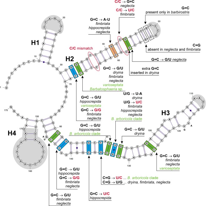
Thirteen morphologically similar strains of barbatosphaeria- and tectonidula-like fungi were studied based on the comparison of cultural and morphological features of sexual and asexual morphs and phylogenetic analyses of five nuclear loci, i.e. internal transcribed spacer rDNA operon (ITS), large and small subunit nuclear ribosomal DNA, β-tubulin, and second largest subunit of RNA polymerase II. Phylogenetic results were supported by in-depth comparative analyses of common core secondary structure of ITS1 and ITS2 in all strains and the identification of non-conserved, co-evolving nucleotides that maintain base pairing in the RNA transcript. Barbatosphaeria is defined as a well-supported monophyletic clade comprising several lineages and is placed in the Sordariomycetes incertae sedis. The genus is expanded to encompass nine species with both septate and non-septate ascospores in clavate, stipitate asci with a non-amyloid apical annulus and non-stromatic ascomata with a long decumbent neck and carbonised wall often covered by pubescence. The asexual morphs are dematiaceous hyphomycetes with holoblastic conidiogenesis belonging to Ramichloridium and Sporothrix types. The morphologically similar Tectonidula, represented by the type species T. hippocrepida, grouped with members of Barbatosphaeria and is transferred to that genus. Four new species are introduced and three new combinations in Barbatosphaeria are proposed. A dichotomous key to species accepted in the genus is provided.
Keywords: Ramichloridium; Sporothrix; Tectonidula; phylogenetics; sequence analysis; spacer regions.
Figures


Similar articles
-
New fungal genera, Tectonidula gen. nov. for Calosphaeria-like fungi with holoblastic-denticulate conidiogenesis and Natantiella gen. nov. for three species segregated from Ceratostomella.Mycol Res. 2009 Sep;113(Pt 9):991-1002. doi: 10.1016/j.mycres.2009.06.003. Epub 2009 Jun 17. Mycol Res. 2009. PMID: 19539759
-
Novel evolutionary lineages revealed in the Chaetothyriales (fungi) based on multigene phylogenetic analyses and comparison of its secondary structure.PLoS One. 2013 May 28;8(5):e63547. doi: 10.1371/journal.pone.0063547. Print 2013. PLoS One. 2013. PMID: 23723988 Free PMC article.
-
Introducing the Rhamphoriaceae, fam. nov. (Sordariomycetes), two new genera, and new life histories for taxa with Phaeoisaria- and Idriella-like anamorphs.Mycologia. 2018 Jul-Aug;110(4):750-770. doi: 10.1080/00275514.2018.1475164. Epub 2018 Aug 20. Mycologia. 2018. PMID: 30125239
-
Phylogenetic classification and generic delineation of Calyptosphaeria gen. nov., Lentomitella, Spadicoides and Torrentispora (Sordariomycetes).Stud Mycol. 2018 Mar;89:1-62. doi: 10.1016/j.simyco.2017.11.004. Epub 2017 Dec 6. Stud Mycol. 2018. PMID: 29367793 Free PMC article.
-
Phylogenetic classification of Pleurothecium and Pleurotheciella gen. nov. and its dactylaria-like anamorph (Sordariomycetes) based on nuclear ribosomal and protein-coding genes.Mycologia. 2012 Nov-Dec;104(6):1299-314. doi: 10.3852/12-035. Epub 2012 Jun 8. Mycologia. 2012. PMID: 22684295
Cited by
-
Compensatory Base Changes in ITS2 Secondary Structure Alignment, Modelling, and Molecular Phylogeny: An Integrated Approach to Improve Species Delimitation in Tulasnella (Basidiomycota).J Fungi (Basel). 2023 Aug 31;9(9):894. doi: 10.3390/jof9090894. J Fungi (Basel). 2023. PMID: 37755002 Free PMC article.
-
Phylogeny, Global Biogeography and Pleomorphism of Zanclospora.Microorganisms. 2021 Mar 29;9(4):706. doi: 10.3390/microorganisms9040706. Microorganisms. 2021. PMID: 33805574 Free PMC article.
-
A new genus and species of Phialocephala-like fungi with cordiform conidia in Chaetosphaeriaceae.Mycoscience. 2024 Nov 27;66(1):109-115. doi: 10.47371/mycosci.2024.11.001. eCollection 2025. Mycoscience. 2024. PMID: 40672143 Free PMC article.
-
Thyridium revised: Synonymisation of Phialemoniopsis under Thyridium and establishment of a new order, Thyridiales.MycoKeys. 2022 Feb 1;86:147-176. doi: 10.3897/mycokeys.86.78989. eCollection 2022. MycoKeys. 2022. PMID: 35145340 Free PMC article.
-
Re-evaluation of Ceratostomella and Xylomelasma with introduction of two new species (Sordariomycetes).MycoKeys. 2024 Nov 21;110:319-360. doi: 10.3897/mycokeys.110.136844. eCollection 2024. MycoKeys. 2024. PMID: 39619666 Free PMC article.
References
-
- Amato A, Kooistra WHCF, Ghiron JHL, et al. 2007. Reproductive isolation among sympatric cryptic species in marine diatoms. Protist 158: 193–207. - PubMed
-
- Bridge PD, Schlitt T, Cannon PF, et al. 2008. Domain II hairpin structure in ITS1 sequences as an aid in differentiating recently evolved animal and plant pathogenic fungi. Mycopathologia 166: 1–16. - PubMed
-
- Campbell J, Shearer CA. 2004. Annulusmagnus and Ascitendus, two new genera in the Annulatascaceae. Mycologia 96: 822–833. - PubMed
LinkOut - more resources
Full Text Sources
Molecular Biology Databases