

Increased synapsin I expression in cerebral malaria
- PMID: 26823711
- PMCID: PMC4713497
Increased synapsin I expression in cerebral malaria
Abstract
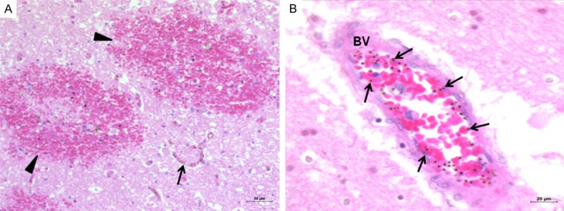
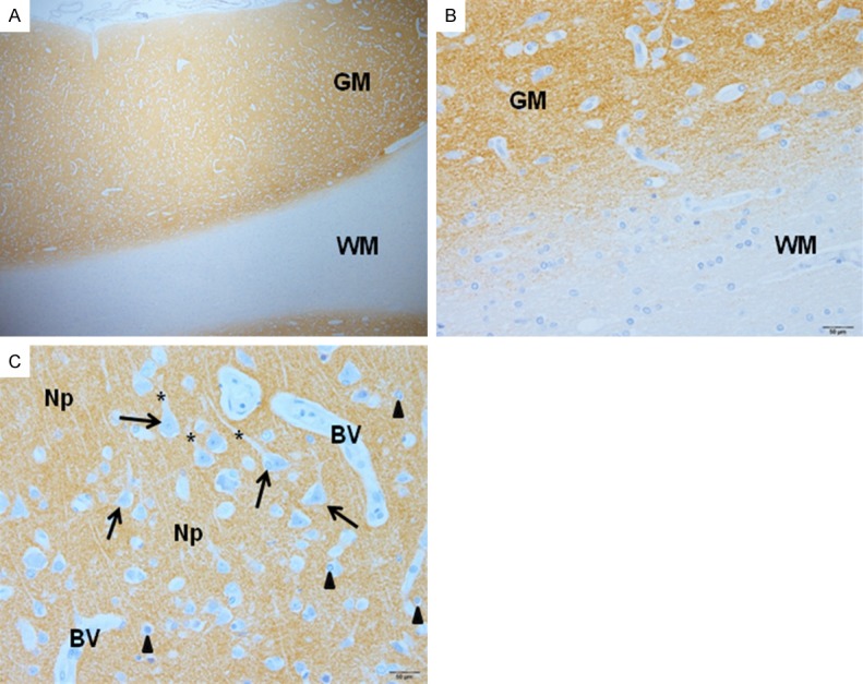
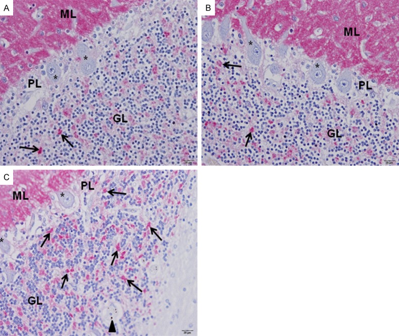
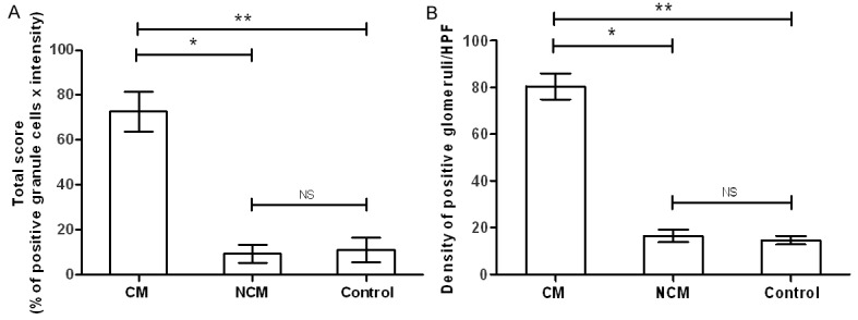
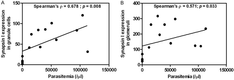
Synapsin I is a neuronal phosphoprotein contained in the synaptic vesicles of mammalian central and peripheral nervous systems. It regulates both neurotransmitter release and synaptic formation. Variations in synapsin I expression in the brain have been reported to cause brain malfunction. In severe malaria, neurological complications, such as convulsion, delirium and coma, suggest abnormalities in the release of neurotransmitters. This study evaluated synapsin I expression in cerebral malaria (CM). An immunohistochemical method was used to study the semi-quantitative and qualitative expression of synapsin I in the brain of CM patients (10 cases) who died with Plasmodium falciparum, compared with non-cerebral malaria (NCM) (4 cases), and control brain tissues (5). Synapsin I was expressed in the gray matter of the cerebral cortex and the molecular layer of the cerebellum, as a diffusely dense precipitate pattern in the neuropil, with no immunoreactivity in the neurons, neuronal dendrites, glial cells, endothelial cells, and Purkinje cells. The findings were similarly demonstrated in CM, NCM, and control brain tissues. However, in the granular layer of the cerebellum, a significant increase in synapsin I expression was observed in the granule cells, and the glomerular synaptic complex, from the CM group, compared with the NCM, and control brain tissues (all P < 0.05). Parasitemia showed a positive correlation with synapsin I expression in the granule cells (on admission: Spearman's ρ = 0.600, P = 0.023) (before death: Spearman's ρ = 0.678, P = 0.008), and glomerular synaptic complex (before death: Spearman's ρ = 0.571, P = 0.033). It was hypothesized that CM causes pre-synaptic excitation and eventually activation of synapsin I, leading to increased neurotransmitter release. Synapsin I inhibitor should be investigated further as a target for a therapeutic intervention to alleviate neurological symptoms in severe malaria.
Keywords: Malaria; Plasmodium falciparum; cerebral malaria; immunohistochemistry; synapsin I.
Figures

Similar articles
-
An ultrastructural study of the brain in fatal Plasmodium falciparum malaria.Am J Trop Med Hyg. 2003 Oct;69(4):345-59. Am J Trop Med Hyg. 2003. PMID: 14640492
-
Genetic analysis of circulating and sequestered populations of Plasmodium falciparum in fatal pediatric malaria.J Infect Dis. 2006 Jul 1;194(1):115-22. doi: 10.1086/504689. Epub 2006 May 31. J Infect Dis. 2006. PMID: 16741890
-
Quantitative Assessment of Multiorgan Sequestration of Parasites in Fatal Pediatric Cerebral Malaria.J Infect Dis. 2015 Oct 15;212(8):1317-21. doi: 10.1093/infdis/jiv205. Epub 2015 Apr 7. J Infect Dis. 2015. PMID: 25852120 Free PMC article.
-
Cerebral Malaria and Neuronal Implications of Plasmodium Falciparum Infection: From Mechanisms to Advanced Models.Adv Sci (Weinh). 2022 Dec;9(36):e2202944. doi: 10.1002/advs.202202944. Epub 2022 Oct 27. Adv Sci (Weinh). 2022. PMID: 36300890 Free PMC article. Review.
-
Cerebral manifestations of falciparum malaria in adults: more than meets the eye.Trends Parasitol. 2025 Apr;41(4):271-279. doi: 10.1016/j.pt.2025.02.005. Epub 2025 Mar 10. Trends Parasitol. 2025. PMID: 40068979 Free PMC article. Review.
Cited by
-
Perturbation of synapsins homeostasis through HIV-1 Tat-mediated suppression of BAG3 in primary neuronal cells.Cell Death Dis. 2019 Jun 17;10(7):473. doi: 10.1038/s41419-019-1702-2. Cell Death Dis. 2019. PMID: 31209204 Free PMC article.
-
Infectious disease-associated encephalopathies.Crit Care. 2021 Jul 6;25(1):236. doi: 10.1186/s13054-021-03659-6. Crit Care. 2021. PMID: 34229735 Free PMC article. Review.
-
The spectrum of clinical biomarkers in severe malaria and new avenues for exploration.Virulence. 2022 Dec;13(1):634-653. doi: 10.1080/21505594.2022.2056966. Virulence. 2022. PMID: 36036460 Free PMC article. Review.
References
-
- Severe falciparum malaria. World Health Organization, Communicable Diseases Cluster. Trans R Soc Trop Med Hyg. 2000;94(Suppl 1):S1–90. - PubMed
-
- Miranda AS, Vieira LB, Lacerda-Queiroz N, Souza AH, Rodrigues DH, Vilela MC, Gomez MV, Machado FS, Rachid MA, Teixeira AL. Increased levels of glutamate in the central nervous system are associated with behavioral symptoms in experimental malaria. Braz J Med Biol Res. 2010;43:1173–1177. - PubMed
-
- Idro R, Jenkins NE, Newton CR. Pathogenesis, clinical features, and neurological outcome of cerebral malaria. Lancet Neurol. 2005;4:827–840. - PubMed
-
- Medana IM, Chaudhri G, Chan-Ling T, Hunt NH. Central nervous system in cerebral malaria: ‘Innocent bystander’ or active participant in the induction of immunopathology? Immunol Cell Biol. 2001;79:101–120. - PubMed
Publication types
MeSH terms
Substances
LinkOut - more resources
Full Text Sources