

Primary splenic angiosarcoma with fever and anemia: a case report and literature review
- PMID: 26823717
- PMCID: PMC4713503
Primary splenic angiosarcoma with fever and anemia: a case report and literature review
Abstract
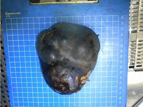
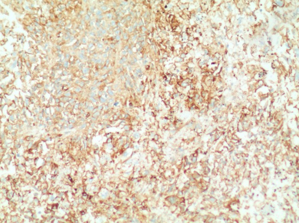
Primary splenic angiosarcoma is an extremely rare and aggressive neoplasm. The prognosis of this disease is dismal, and the mean survival is less than 6 months after the diagnosis. This neoplasm typically presents with abdominal pain, splenomegaly, weight loss, and spontaneous splenic rupture. Fever is a very rare presentation of splenic angiosarcoma. Here we report the case of a 64-year-old man who presented with fever and anemia. A laparoscopic splenectomy was performed and revealed splenic angiosarcoma. The postoperative course was uneventful and the patient received 5 cycles of adjuvant chemotherapy with ifosfamide plus epirubicin. He remained disease free at 9 months after surgery. This is the first case of splenic angiosarcoma with fever as the initial presentation that was treated with laparoscopic splenectomy to be reported in the English literature.
Keywords: Splenic angiosarcoma; anemia; disease free; fever; splenectomy.
Figures


Similar articles
-
[Spontaneous spleen rupture due to primary splenic angiosarcoma: a case report].Ulus Travma Acil Cerrahi Derg. 2007 Oct;13(4):313-5. Ulus Travma Acil Cerrahi Derg. 2007. PMID: 17978914 Turkish.
-
Primary splenic angiosarcoma.JSLS. 2010 Jul-Sep;14(3):431-5. doi: 10.4293/108680810X12924466006521. JSLS. 2010. PMID: 21333203 Free PMC article.
-
Primary angiosarcoma of the spleen.J Surg Oncol. 2005 Dec 15;92(4):312-6. doi: 10.1002/jso.20419. J Surg Oncol. 2005. PMID: 16299797
-
Case report of primary splenic angiosarcoma with hepatic metastases.World J Gastroenterol. 2015 Oct 21;21(39):11199-204. doi: 10.3748/wjg.v21.i39.11199. World J Gastroenterol. 2015. PMID: 26494974 Free PMC article. Review.
-
[Rupture of the spleen in angiosarcoma: a case report and review of the literature].Chir Ital. 2005 May-Jun;57(3):377-80. Chir Ital. 2005. PMID: 16231829 Review. Italian.
Cited by
-
Primary splenic angiosarcoma: a rare entity often associated with rupture and hemoperitoneum.Autops Case Rep. 2019 Jul 12;9(3):e2019100. doi: 10.4322/acr.2019.100. eCollection 2019 Jul-Sep. Autops Case Rep. 2019. PMID: 31372360 Free PMC article.
-
Primary Colonic Angiosarcoma Seen in a Patient on Calcium Channel Blocker: A Case Report with Summary Analysis of 32 Other Cases from the Literature.Am J Case Rep. 2018 Mar 7;19:254-261. doi: 10.12659/ajcr.907287. Am J Case Rep. 2018. PMID: 29511155 Free PMC article. Review.
-
Pancytopenia Related to Splenic Angiosarcoma: A Case Report and Literature Review.Hematol Rep. 2024 Oct 18;16(4):648-655. doi: 10.3390/hematolrep16040063. Hematol Rep. 2024. PMID: 39449306 Free PMC article.
-
A primary splenic angiosarcoma hepatic metastasis after splenectomy and its genomic alteration profile.Medicine (Baltimore). 2019 Jul;98(28):e16245. doi: 10.1097/MD.0000000000016245. Medicine (Baltimore). 2019. PMID: 31305404 Free PMC article.
References
-
- Neuhauser TS, Derringer GA, Thompson LD, Fanburg-Smith JC, Miettinen M, Saaristo A, Abbondanzo SL. Splenic angiosarcoma: a clinicopathologic and immunophenotypic study of 28 cases. Mod Pathol. 2000;13:978–987. - PubMed
-
- Falk S, Krishnan J, Meis JM. Primary angiosarcoma of the spleen. A clinicopathologic study of 40 cases. Am J Surg Pathol. 1993;17:959–970. - PubMed
-
- Kren L, Kaur P, Goncharuk VN, Dolezel Z, Krenova Z. Primary angiosarcoma of the spleen in a child. Med Pediatr Oncol. 2003;40:411–412. - PubMed
Publication types
MeSH terms
Substances
LinkOut - more resources
Full Text Sources
Medical