

Procoagulant role of neutrophil extracellular traps in patients with gastric cancer
- PMID: 26823721
- PMCID: PMC4713507
Procoagulant role of neutrophil extracellular traps in patients with gastric cancer
Abstract
Background: Patients with gastric cancer (GC) commonly exhibit a hypercoagulable state that results in significant morbidity and mortality. Recent studies have shown that neutrophil extracellular traps (NETs) trigger coagulation through an intrinsic pathway and contribute to thrombus initiation and progression. In this study, we aimed to determine the procoagulant activity (PCA) of NETs in patients with GC.
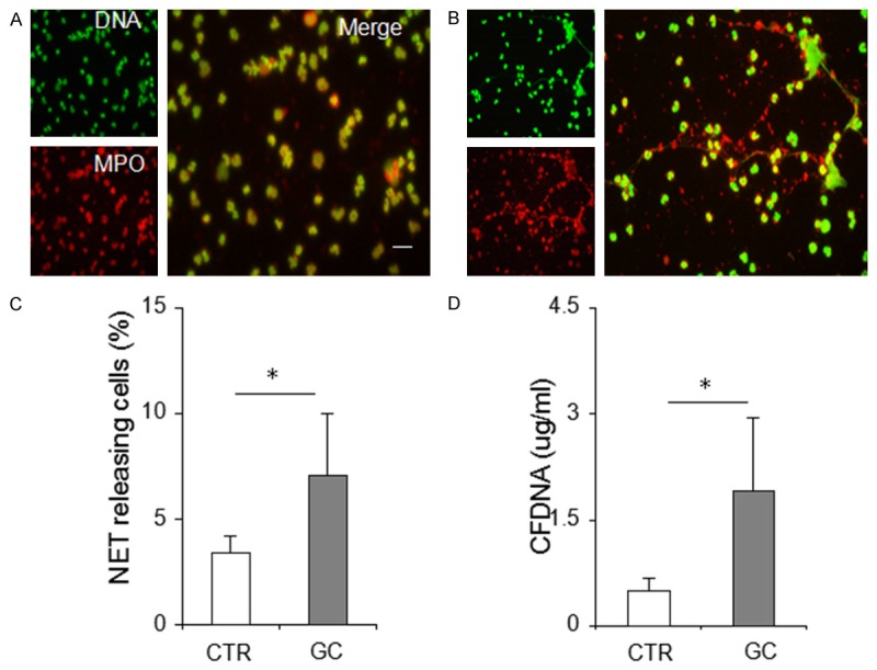
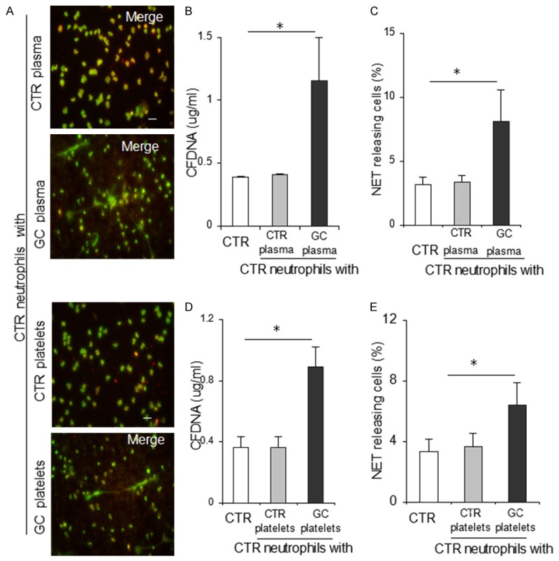
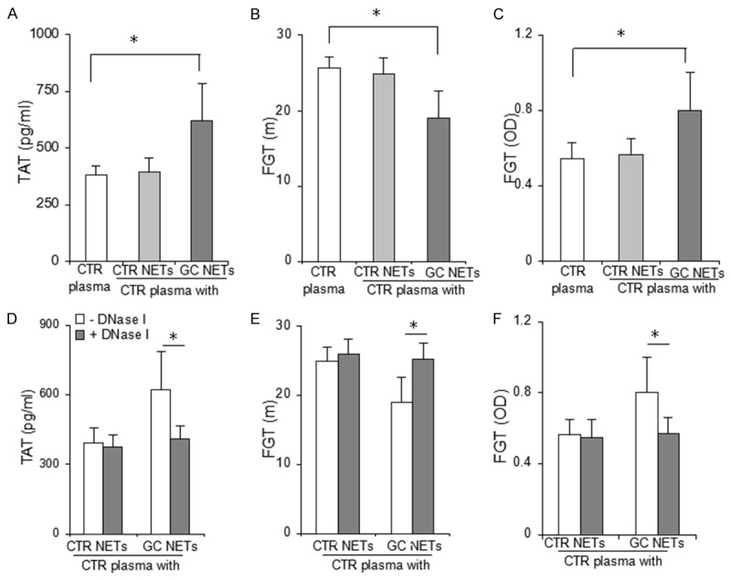
Methods: NET formation and their PCAs were assessed in 48 patients with GC and 36 healthy controls using immunofluorescence microscopy of neutrophil markers and extracellular DNA as well as a modified capture ELISA technique, and thrombin-antithrombin complex and clot (fibrin) spectroscopic detection, respectively.
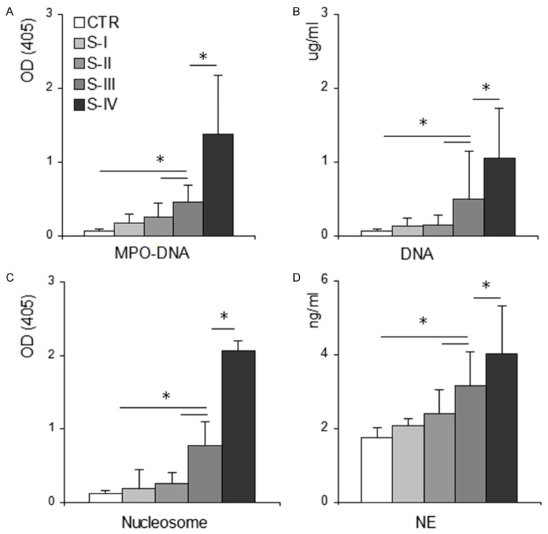
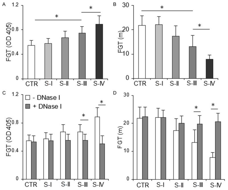
Results: Here we showed that neutrophils isolated from patients with GC displayed significantly enhanced NET formation compared with those from healthy controls; furthermore, plasma or platelets obtained from patients with GC induced control neutrophils to release NETs. In addition, NETs released by GC neutrophils significantly increased the potency of control plasma to generate thrombin and fibrin. Notably, these procoagulant effects were dramatically attenuated by application of DNase I. We further found that spontaneous NET formation in patients with GC was significantly higher than that in controls, increased with tumor- node-metastasis stage elevation, and positively correlated with thrombin-antithrombin complex levels and D-dimers. Additionally, the effect of DNase I on cell-free plasma generation of fibrin was dependent on the concentration of NET formation.
Conclusion: These results suggest that GC creates a systemic environment that primes neutrophils to release procoagulant NETs. Thus, targeting NETs might improve the coagulopathy of patients with GC.
Keywords: NETs; Stomach neoplasm; cell-free DNA; neutrophils; prothrombotic state.
Figures
Similar articles
-
Neutrophil extracellular traps participate in the development of cancer-associated thrombosis in patients with gastric cancer.World J Gastroenterol. 2022 Jul 14;28(26):3132-3149. doi: 10.3748/wjg.v28.i26.3132. World J Gastroenterol. 2022. PMID: 36051331 Free PMC article.
-
Neutrophil Extracellular Traps Promote Hypercoagulability in Patients With Sepsis.Shock. 2017 Feb;47(2):132-139. doi: 10.1097/SHK.0000000000000741. Shock. 2017. PMID: 27617671
-
Phosphotidylserine exposure and neutrophil extracellular traps enhance procoagulant activity in patients with inflammatory bowel disease.Thromb Haemost. 2016 Apr;115(4):738-51. doi: 10.1160/TH15-09-0710. Epub 2015 Dec 10. Thromb Haemost. 2016. PMID: 26660948
-
The pathway of neutrophil extracellular traps towards atherosclerosis and thrombosis.Atherosclerosis. 2019 Sep;288:9-16. doi: 10.1016/j.atherosclerosis.2019.06.919. Epub 2019 Jun 29. Atherosclerosis. 2019. PMID: 31280097 Review.
-
Neutrophil Extracellular Traps: Villains and Targets in Arterial, Venous, and Cancer-Associated Thrombosis.Arterioscler Thromb Vasc Biol. 2019 Sep;39(9):1724-1738. doi: 10.1161/ATVBAHA.119.312463. Epub 2019 Jul 18. Arterioscler Thromb Vasc Biol. 2019. PMID: 31315434 Free PMC article. Review.
Cited by
-
The significance of coagulation and fibrinolysis-related parameters in predicting postoperative venous thrombosis in patients with breast cancer.Gland Surg. 2021 Apr;10(4):1439-1446. doi: 10.21037/gs-21-117. Gland Surg. 2021. PMID: 33968695 Free PMC article.
-
Neutrophil extracellular traps participate in the development of cancer-associated thrombosis in patients with gastric cancer.World J Gastroenterol. 2022 Jul 14;28(26):3132-3149. doi: 10.3748/wjg.v28.i26.3132. World J Gastroenterol. 2022. PMID: 36051331 Free PMC article.
-
Cancers Cells in Traps? The Pathways of NETs Formation in Response to OSCC in Humans-A Pilot Study.Cancer Control. 2020 Jan-Dec;27(1):1073274820960473. doi: 10.1177/1073274820960473. Cancer Control. 2020. PMID: 33073595 Free PMC article.
-
Neutrophil extracellular traps promote gastric cancer metastasis by inducing epithelial‑mesenchymal transition.Int J Mol Med. 2021 Jul;48(1):127. doi: 10.3892/ijmm.2021.4960. Epub 2021 May 20. Int J Mol Med. 2021. PMID: 34013374 Free PMC article.
-
Neutrophil Extracellular Traps in Digestive Cancers: Warrior or Accomplice.Front Oncol. 2021 Nov 19;11:766636. doi: 10.3389/fonc.2021.766636. eCollection 2021. Front Oncol. 2021. PMID: 34868992 Free PMC article. Review.
References
-
- Di Micco P, Romano M, Niglio A, Nozzolillo P, Federico A, Petronella P, Nunziata L, Di Micco B, Torella R. Alteration of haemostasis in nonmetastatic gastric cancer. Dig Liver Dis. 2001;33:546–50. - PubMed
-
- Fidan E, Kavgaci H, Orem A, Yilmaz M, Yildiz B, Fidan S, Akcan B, Ozdemir F, Aydin F. Thrombin activatable fibrinolysis inhibitor and thrombinantithrombin-III-complex levels in patients with gastric cancer. Tumour Biol. 2012;33:1519–25. - PubMed
-
- Kwon HC, Oh SY, Lee S, Kim SH, Han JY, Koh RY, Kim MC, Kim HJ. Plasma levels of prothrombin fragment F1+2, D-dimer and prothrombin time correlate with clinical stage and lymph node metastasis in operable gastric cancer patients. Jpn J Clin Oncol. 2008;38:2–7. - PubMed
-
- Young A, Chapman O, Connor C, Poole C, Rose P, Kakkar AK. Thrombosis and cancer. Nat Rev Clin Oncol. 2012;9:437–49. - PubMed
-
- Timp JF, Braekkan SK, Versteeg HH, Cannegieter SC. Epidemiology of cancer-associated venous thrombosis. Blood. 2013;122:1712–23. - PubMed
Publication types
MeSH terms
Substances
LinkOut - more resources
Full Text Sources
Medical
Miscellaneous