

Methylation-silencing RCC1 expression is associated with tumorigenesis and depth of invasion in gastric cancer
- PMID: 26823742
- PMCID: PMC4713528
Methylation-silencing RCC1 expression is associated with tumorigenesis and depth of invasion in gastric cancer
Abstract
Introduction: Regulator of chromosome condensation 1 (RCC1) is a critical cell cycle regulator. We firstly identified RCC1 gene hypermethylation in gastric tumor tissues using the differential methylation hybridization (DMH) microarray, but the role of RCC1 in the pathogenesis of gastric carcinoma is largely unknown.
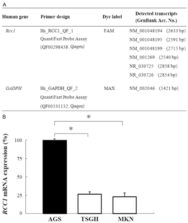
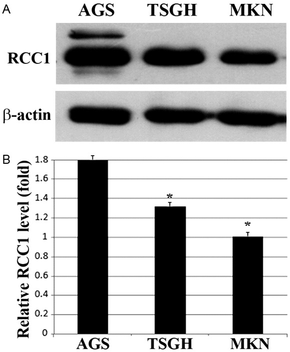
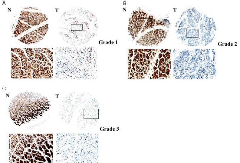
Methods: Three gastric cancer cell lines (AGS, MKN45, and TSGH9201) were used to analyze RCC1 gene methylation, mRNA and protein expressions. Furthermore, 85 pairs of matched human gastric carcinoma samples in a tissue microarray were used to analyze RCC1 expression by immunohistochemistry staining.
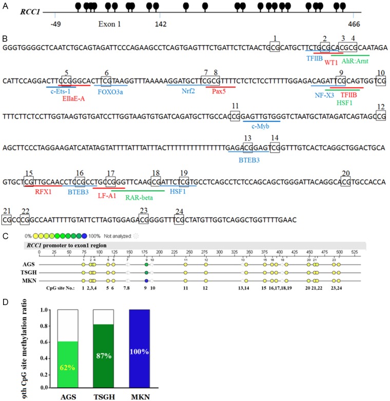
Results: A differential methylation pattern was found in TSGH9201 (100%), MKN45 (87%), and AGS (62%) cell lines at the 9th CpG site of RCC1 exon 1. RCC1 mRNA and protein expressions in AGS cells were significantly higher than in TSGH9201 and MKN45 cell lines (P < 0.05). Tissue array data showed that RCC1 expression was detected in 21% (18/85) of gastric carcinoma tissues and in 80% (76/95) of adjacent non-tumor tissues. The expression of RCC1 in gastric carcinoma tissues was significantly lower than in adjacent non-tumor tissues (P < 0.001). Furthermore, an association between RCC1 expression and clinicopathological features showed that RCC1 expression was closely correlated with tumor differentiation and depth of invasion (P < 0.05).
Conclusions: Our data indicate that RCC1 expression is frequently lost in poorly differentiated gastric cell lines and gastric carcinoma tissues. Loss of RCC1 expression is correlated with tumor differentiation and depth of invasion. These findings suggest that RCC1 may play a tumor suppressor role in gastric carcinoma.
Keywords: DMH microarray; DNA methylation; RCC1; gastric carcinoma; immunohistochemistry; invasion; tumor tissue array.
Figures
Similar articles
-
KRAB zinc-finger protein 382 regulates epithelial-mesenchymal transition and functions as a tumor suppressor, but is silenced by CpG methylation in gastric cancer.Int J Oncol. 2018 Sep;53(3):961-972. doi: 10.3892/ijo.2018.4446. Epub 2018 Jun 19. Int J Oncol. 2018. PMID: 29956735 Free PMC article.
-
CpG island promoter hypermethylation of Ras association domain family 1A gene contributes to gastric carcinogenesis.Mol Med Rep. 2015 Apr;11(4):3039-46. doi: 10.3892/mmr.2014.3055. Epub 2014 Dec 5. Mol Med Rep. 2015. PMID: 25483734
-
Zinc-finger protein 331, a novel putative tumor suppressor, suppresses growth and invasiveness of gastric cancer.Oncogene. 2013 Jan 17;32(3):307-17. doi: 10.1038/onc.2012.54. Epub 2012 Feb 27. Oncogene. 2013. PMID: 22370639
-
Recent progress in the study of methylated tumor suppressor genes in gastric cancer.Chin J Cancer. 2013 Jan;32(1):31-41. doi: 10.5732/cjc.011.10175. Epub 2011 Nov 4. Chin J Cancer. 2013. PMID: 22059906 Free PMC article. Review.
-
The intricate roles of RCC1 in normal cells and cancer cells.Biochem Soc Trans. 2022 Feb 28;50(1):83-93. doi: 10.1042/BST20210861. Biochem Soc Trans. 2022. PMID: 35191966 Review.
Cited by
-
Retinol-binding protein type 1 expression predicts poor prognosis in head and neck squamous cell carcinoma.BMC Cancer. 2024 Oct 15;24(1):1277. doi: 10.1186/s12885-024-12565-3. BMC Cancer. 2024. PMID: 39407127 Free PMC article.
-
Villi-specific Gene Expression Reveals Novel Prognostic Biomarkers in Multiple Human Cancers.J Cancer. 2017 Aug 23;8(14):2793-2801. doi: 10.7150/jca.19787. eCollection 2017. J Cancer. 2017. PMID: 28928868 Free PMC article.
-
RCC1 Expression as a Prognostic Marker in Colorectal Liver Oligometastases.Pathol Oncol Res. 2021 Dec 2;27:1610077. doi: 10.3389/pore.2021.1610077. eCollection 2021. Pathol Oncol Res. 2021. PMID: 34924821 Free PMC article.
-
Decoding oncogenic secrets of regulator of chromosome condensation 1: A breakthrough mechanistic evidence from breast and lung cancer models.PLoS One. 2025 Mar 31;20(3):e0319748. doi: 10.1371/journal.pone.0319748. eCollection 2025. PLoS One. 2025. PMID: 40163507 Free PMC article.
-
Regulator of chromatin condensation 1 abrogates the G1 cell cycle checkpoint via Cdk1 in human papillomavirus E7-expressing epithelium and cervical cancer cells.Cell Death Dis. 2018 May 22;9(6):583. doi: 10.1038/s41419-018-0584-z. Cell Death Dis. 2018. PMID: 29789527 Free PMC article.
References
-
- Bertuccio P, Chatenoud L, Livi F, Proud D, Ferlay J, Negri E, Malvezzi M, La Vecchia C. Recent pattern in gastric cancer: a global overview. Int J Cancer. 2009;125:666–673. - PubMed
-
- Brenner H, Rothenbacher D, Amdt V. Epidemiology of stomach cancer. Methods Mol Biol. 2009;472:467–477. - PubMed
-
- Ferlay J, Shin HR, Bray F, Forman D, Mathers C, Parkin DM. Estimates of worldwide burden of cancer in 2008: GLOBOCAN 2008. Int J Cancer. 2010;127:2893–2917. - PubMed
-
- Leung WK, Wu MS, Kakugawa Y, Kim JJ, Yeoh KG, Goh KL, Wu KC, Wu DC, Sollano J, Kachintorn U, Gotoda T, Lin JT, You WC, Ng EK, Sung JJ. Screening for gastric cancer in Asia: current evidence and practice. Lancet Oncol. 2008;9:279–287. - PubMed
Publication types
MeSH terms
Substances
LinkOut - more resources
Full Text Sources
Other Literature Sources
Medical
Research Materials