

Lobulated adenomyoepithelioma: a case report showing immunohistochemical profiles
- PMID: 26823903
- PMCID: PMC4713689
Lobulated adenomyoepithelioma: a case report showing immunohistochemical profiles
Abstract
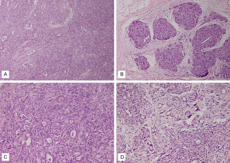
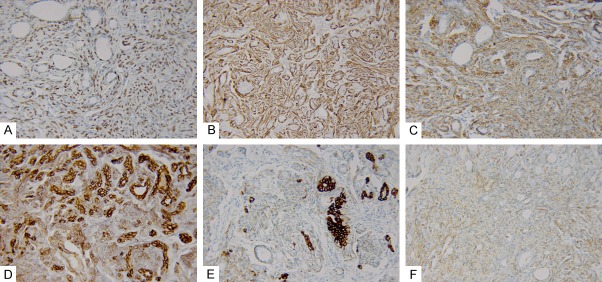
Lobulated adenomyoepithelioma of the breast is an extremely rare lesion, with hyperplasia of myoepithelial cells and glandular epithelial cells. We present a case of a 51-year-old woman with a small painless hard lump in each breast. The lesion in the left breast was an irregular solid mass, and the right breast showed a subareolar nodule with bloodstained nipple discharge. The final diagnosis was intraductal papillary carcinoma in the right breast and lobulated adenomyoepithelioma in the left breast. In the left breast lesion, histopathologic examination revealed multiple nodules composed of proliferative glandular epithelial cells and surrounding myoepithelial cells. Solid nests of clear or eosinophilic myoepithelial cells proliferated around compressed epithelial-lined space. Smaller satellite nodules were seen. Immunohistochemistry revealed myoepithelial cells were positive for P63, smooth muscle actin, calponin, 34βE12, CK5/6 and CK14, while glandular epithelial cells were positive for AE1/AE3 and CK7. Lobulated adenomyoepithelioma has a high chance of recurrence and malignant degeneration due to inadequate excision. Therefore, understanding of the pathological morphology and accurate diagnosis is important for surgical planning. Moreover, close follow-up is recommended for patients with lobulated adenomyoepithelioma despite the lesion being reported as benign.
Keywords: Adenomyoepithelioma; breast; lobulated; myoepithelium.
Figures
References
-
- Tavassoli FA. Myoepithelial lesions of the breast. Myoepitheliosis, adenomyoepithelioma, and myoepithelial carcinoma. Am J Surg Pathol. 1991;15:554–568. - PubMed
-
- Kiaer H, Nielsen B, Paulsen S, Sorensen IM, Dyreborg U, Blichert-Toft M. Adenomyoepithelial adenosis and low-grade malignant adenomyoepithelioma of the breast. Virchows Arch A Pathol Anat Histopathol. 1984;405:55–67. - PubMed
-
- Tsuda H, Mukai K, Fukutomi T, Hirohashi S. Malignant progression of adenomyoepithelial adenosis of the breast. Pathol Int. 1994;44:475–479. - PubMed
-
- Tavassoli F, Soares J, editors. Myoepithelial Lesion: World Health Organization classification of Tumors. Pathology & Genetics. Tumors of the breast and Female Genital Organs. Lyon: International Agency for Research on Cancer (IARC); 2003. pp. 98–102.
-
- Schmitt F, Tan P, Dabbs D, Jones L, editors. Myoepithelial and epithelialmyoepithelial lesions: World Health Organization classification of Tumors of the breast. Lyon: International Agency for Research on Cancer (IARC); 2012. pp. 119–122.
Publication types
MeSH terms
Substances
LinkOut - more resources
Full Text Sources
Medical
Research Materials
Miscellaneous