Altered functional connectivity of the amygdaloid input nuclei in adolescents and young adults with autism spectrum disorder: a resting state fMRI study
- PMID: 26823966
- PMCID: PMC4730628
- DOI: 10.1186/s13229-015-0060-x
Altered functional connectivity of the amygdaloid input nuclei in adolescents and young adults with autism spectrum disorder: a resting state fMRI study
Abstract
Background: Amygdala dysfunction is hypothesized to underlie the social deficits observed in autism spectrum disorders (ASD). However, the neurobiological basis of this hypothesis is underspecified because it is unknown whether ASD relates to abnormalities of the amygdaloid input or output nuclei. Here, we investigated the functional connectivity of the amygdaloid social-perceptual input nuclei and emotion-regulation output nuclei in ASD versus controls.
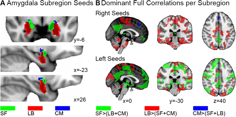
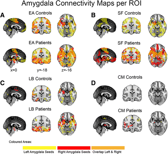
Methods: We collected resting state functional magnetic resonance imaging (fMRI) data, tailored to provide optimal sensitivity in the amygdala as well as the neocortex, in 20 adolescents and young adults with ASD and 25 matched controls. We performed a regular correlation analysis between the entire amygdala (EA) and the whole brain and used a partial correlation analysis to investigate whole-brain functional connectivity uniquely related to each of the amygdaloid subregions.
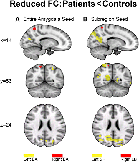
Results: Between-group comparison of regular EA correlations showed significantly reduced connectivity in visuospatial and superior parietal areas in ASD compared to controls. Partial correlation analysis revealed that this effect was driven by the left superficial and right laterobasal input subregions, but not the centromedial output nuclei.
Conclusions: These results indicate reduced connectivity of specifically the amygdaloid sensory input channels in ASD, suggesting that abnormal amygdalo-cortical connectivity can be traced down to the socio-perceptual pathways.
Keywords: Amygdala; Autism spectrum disorder; Centromedial; Connectivity; Input-output; Laterobasal; Nuclei; Social perception; Superficial.
Figures


Similar articles
-
Altered functional organization within the insular cortex in adult males with high-functioning autism spectrum disorder: evidence from connectivity-based parcellation.Mol Autism. 2016 Oct 5;7:41. doi: 10.1186/s13229-016-0106-8. eCollection 2016. Mol Autism. 2016. PMID: 27713815 Free PMC article.
-
Subregional differences in intrinsic amygdala hyperconnectivity and hypoconnectivity in autism spectrum disorder.Autism Res. 2016 Jul;9(7):760-72. doi: 10.1002/aur.1589. Epub 2015 Dec 15. Autism Res. 2016. PMID: 26666502 Free PMC article.
-
Somatosensory Regions Show Limited Functional Connectivity Differences in Youth with Autism Spectrum Disorder.Brain Connect. 2018 Nov;8(9):558-566. doi: 10.1089/brain.2018.0614. Brain Connect. 2018. PMID: 30411970
-
Disrupted cortical connectivity theory as an explanatory model for autism spectrum disorders.Phys Life Rev. 2011 Dec;8(4):410-37. doi: 10.1016/j.plrev.2011.10.001. Epub 2011 Oct 13. Phys Life Rev. 2011. PMID: 22018722 Review.
-
Resting-state abnormalities of posterior cingulate in autism spectrum disorder.Prog Mol Biol Transl Sci. 2020;173:139-159. doi: 10.1016/bs.pmbts.2020.04.010. Epub 2020 May 4. Prog Mol Biol Transl Sci. 2020. PMID: 32711808 Review.
Cited by
-
Functional connectivity directionality between large-scale resting-state networks across typical and non-typical trajectories in children and adolescence.PLoS One. 2022 Dec 1;17(12):e0276221. doi: 10.1371/journal.pone.0276221. eCollection 2022. PLoS One. 2022. PMID: 36454744 Free PMC article.
-
Neuroinflammatory Gene Expression Alterations in Anterior Cingulate Cortical White and Gray Matter of Males With Autism Spectrum Disorder.Autism Res. 2020 Jun;13(6):870-884. doi: 10.1002/aur.2284. Epub 2020 Mar 4. Autism Res. 2020. PMID: 32129578 Free PMC article.
-
The Association Between Amygdala Subfield-Related Functional Connectivity and Stigma Reduction 12 Months After Social Contacts: A Functional Neuroimaging Study in a Subgroup of a Randomized Controlled Trial.Front Hum Neurosci. 2020 Aug 27;14:356. doi: 10.3389/fnhum.2020.00356. eCollection 2020. Front Hum Neurosci. 2020. PMID: 33192379 Free PMC article.
-
RDoC-based categorization of amygdala functions and its implications in autism.Neurosci Biobehav Rev. 2018 Jul;90:115-129. doi: 10.1016/j.neubiorev.2018.04.007. Epub 2018 Apr 13. Neurosci Biobehav Rev. 2018. PMID: 29660417 Free PMC article. Review.
-
Abnormal Functional Connectivity of the Primary Sensory Network in Autism Spectrum Disorder: Sex Differences, Early Overdevelopment, and Clinical Significance.Brain Behav. 2025 Mar;15(3):e70363. doi: 10.1002/brb3.70363. Brain Behav. 2025. PMID: 40123151 Free PMC article.
References
-
- American Psychiatric Association . Diagnostic and statistical manual of mental disorders: DSM-IV-TR®. Washington (DC): American Psychiatric Publisher; 2000.
-
- American Psychiatric Association . Diagnostic and statistical manual of mental disorders. 5. Washington (DC): American Psychiatric Publisher; 2013.
Publication types
MeSH terms
LinkOut - more resources
Full Text Sources
Other Literature Sources
Medical

