. 2016 Jan 28:7:9.
doi: 10.1186/s13293-016-0060-3. eCollection 2016.

High-fat diet impairs spatial memory and hippocampal intrinsic excitability and sex-dependently alters circulating insulin and hippocampal insulin sensitivity

Affiliations

High-fat diet impairs spatial memory and hippocampal intrinsic excitability and sex-dependently alters circulating insulin and hippocampal insulin sensitivity

Erica L Underwood et al. Biol Sex Differ. .

Abstract

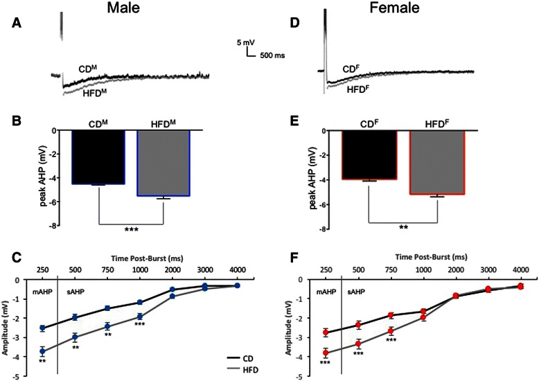
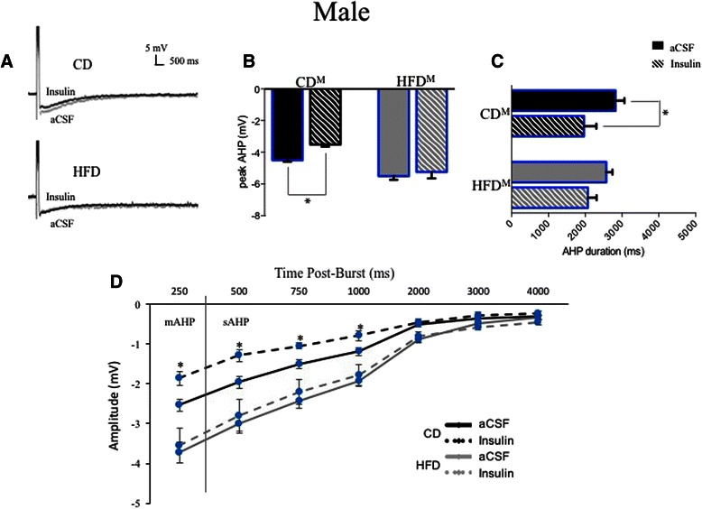
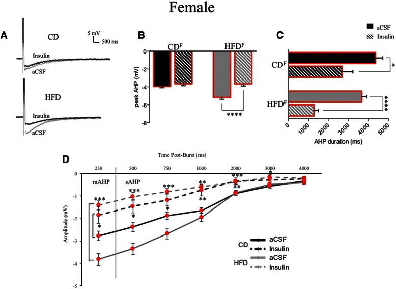
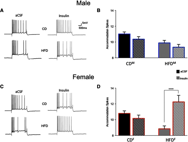
Background: High-fat diets promoting obesity/type-2 diabetes can impair physiology and cognitive performance, although sex-dependent comparisons of these impairments are rarely made. Transient reductions in Ca(2+)-dependent afterhyperpolarizations (AHPs) occur during memory consolidation, enhancing intrinsic excitability of hippocampal CA1 pyramidal neurons. In rats fed standard diets, insulin can enhance memory and reduce amplitude and duration of AHPs.

Methods: Effects of chronic high-fat diet (HFD) on memory, circulating insulin, and neuronal physiology were compared between young adult male and female Long-Evans rats. Rats fed for 12 weeks (from weaning) a HFD or a control diet (CD) were then tested in vivo prior to in vitro recordings from CA1 pyramidal neurons.

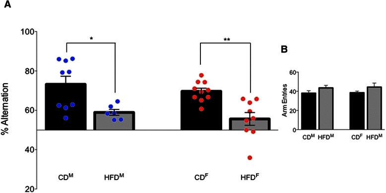
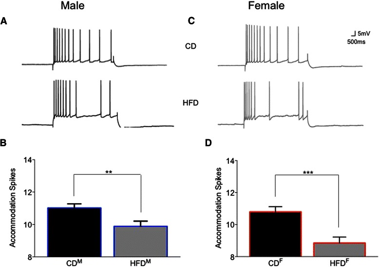
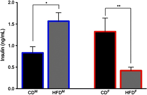
Results: The HFD significantly impaired spatial memory in both males and females. Significant sex differences occurred in circulating insulin and in the insulin sensitivity of hippocampal neurons. Circulating insulin significantly increased in HFD males but decreased in HFD females. While the HFD significantly reduced hippocampal intrinsic excitability in both sexes, CA1 neurons from HFD females remained insulin-sensitive but those from HFD males became insulin-insensitive.

Conclusions: Findings consistent with these have been characterized previously in HFD or senescent males, but the effects observed here in young females are unique. Loss of CA1 neuronal excitability, and sex-dependent loss of insulin sensitivity, can have significant cognitive consequences, over both the short term and the life span. These findings highlight needs for more research into sex-dependent differences, relating systemic and neural plasticity mechanisms in metabolic disorders.

Keywords: AHP; CA1; Diabetes; Glucose regulation; High-fat diet; Hippocampal excitability; Sex differences; Spatial memory.


Figures

Fig. 1
Fig. 1
The spontaneous alternation task used to assess spatial memory. a A four-arm radial plus maze, with two explicit distal visual cues for spatial orientation. No explicit rewards were present on the maze. b The time line used to ensure the behavior of all rats was minimally impacted by stressors related to the environment or the experimenters. c Parsing of exploratory behavior into sets of alternations or non-alternations. d Conversion of raw behavioral observations into % alternation scores for comparisons between individuals and between groups
Fig. 2
Fig. 2
HFD impaired spatial memory on a the spontaneous alternation task in both males and females but did not significantly alter total exploration b of the plus maze (p > 0.2). *p < 0.05; **p < 0.01
Fig. 3
Fig. 3
Ventral CA1 pyramidal neurons showed sex- and diet-dependent differences in intrinsic excitability, here assessed as differences in post-burst AHPs (average traces from male (a) and female (d) neurons are shown). The HFD enhanced peak AHP amplitudes significantly in both males (b) and females (e), but did not significantly increase AHP duration in either sex. No sex differences were observed in measures of medium or slow AHPs from CA1 neurons of control-fed males (c) or females (f). The HFD significantly enhanced both mAHP and sAHP measures in males (c) and more profoundly enhanced mAHP and sAHP components in female neurons (f). **p < 0.01; ***p < 0.001
Fig. 4
Fig. 4
Further evidence of diet-induced reductions in intrinsic excitability is seen in measures of spike-frequency accommodation of ventral CA1 pyramidal neurons. Representative examples of accommodation of male neurons (a) and female neurons (c) from rats fed the two diets for 15 weeks are shown. Significantly increased accommodation (i.e., decreased action potential firing) to a sustained depolarizing pulse was observed in both male (b) and female (d) HFD CA1 neurons. **p < 0.01; ***p < 0.001
Fig. 5
Fig. 5
Sex-dependent effects of the HFD on circulating insulin. In males, the HFD increased circulating insulin compared to controls. However, in females, the HFD significantly decreased circulating insulin compared to controls. While this increased insulin in HFD-fed male rats met a clinical criterion for type-2 diabetes, HFD-fed females not only did not exhibit this clinically relevant profile, but in fact exhibited anomalous decreases in circulating insulin. *p < 0.05; **p < 0.01
Fig. 6
Fig. 6
Sex- and diet-dependent responses to bath application of 12.5 nM insulin in CA1 pyramidal neurons from male rats. a Averaged AHP traces are shown from CD (insulin-sensitive) neurons and HFD (insulin-insensitive) male neurons. b Bath application of insulin significantly reduced peak AHP amplitudes in CD but not HFD neurons. c AHP durations were significantly reduced in CD neurons, but not in HFD neurons. d While mAHP and sAHP measures were significantly reduced by insulin in neurons from CD rats, both mAHPs and sAHPs from HFD rats were insulin-insensitive. *p < 0.05
Fig. 7
Fig. 7
Diet-dependent responses to bath application of 12.5 nM insulin in CA1 pyramidal neurons from female rats. a Averaged AHP traces are shown from both CD neurons and HFD neurons. b Bath application of insulin had no effect on peak AHP amplitudes in CD neurons but significantly reduced peak AHPs in HFD neurons. c While AHP durations were significantly reduced by insulin in CD neurons, significantly larger reductions were observed in HFD neurons. d Both mAHP and sAHP measures were significantly reduced by insulin in neurons from CD rats (black bars at left indicate comparisons made). Notably, insulin produced a more effective reduction of mAHPs and sAHPs in neurons from HFD rats (gray bars at left indicate comparisons made), continuing much longer post-burst, i.e., insulin significantly enhanced intrinsic excitability for many seconds in neurons from female HFD rats. Unlike in males, where HFD neurons lost insulin sensitivity on multiple measures, insulin sensitivity of AHPs actually increased in neurons from HFD female rats. *p < 0.05; **p < 0.01; ***p < 0.001; ****p < 0.0001
Fig. 8
Fig. 8
Sex- and diet-dependent effects of insulin were observed in measures of spike frequency accommodation. In neurons from CD a males and c females, the bath application of insulin did not alter accommodation, with no significant change in firing seen (b, d). Accommodation of HFD male neurons (a) was also unaffected, with no significant change in firing in the presence of insulin (b). In neurons from HFD females, the bath application of insulin (c) actually reduced accommodation, with a significant increase in action potential firing to a sustained depolarizing pulse observed (d). Again, while the HFD did not alter insulin sensitivity in males, it enhanced insulin sensitivity in females. ****p < 0.0001

References

    1. Underwood EL, Thompson LT. A high-fat diet causes impairment in hippocampal memory and sex-dependent alterations in peripheral metabolism. Neural Plast. 2015;501:161985. - PMC - PubMed
    1. Banks WA. The source of cerebral insulin. Eur J Pharmacol. 2004;490:5–12. doi: 10.1016/j.ejphar.2004.02.040. - DOI - PubMed
    1. Peruzzi F, Prisco M, Dews M, Salomoni P, Grassilli E, Romano G, et al. Multiple signaling pathways of the insulin-like growth factor 1 receptor in protection from apoptosis. Mol Cell Biol. 1999;19:7203–15. doi: 10.1128/MCB.19.10.7203. - DOI - PMC - PubMed
    1. Liu L, Brown JC, Webster WW, Morrisett RA, Monaghan DT. Insulin potentiates N-methyl-D-aspartate receptor activity in Xenopus oocytes and rat hippocampus. Neurosci Lett. 1995;192:5–8. doi: 10.1016/0304-3940(95)11593-L. - DOI - PubMed
    1. Rasch B, Büchel C, Gais S, Born J. Odor cues during slow-wave sleep prompt declarative memory consolidation. Science. 2007;315:1426–9. doi: 10.1126/science.1138581. - DOI - PubMed

LinkOut - more resources