A panel of glycoproteins as candidate biomarkers for early diagnosis and treatment evaluation of B-cell acute lymphoblastic leukemia
- PMID: 26823978
- PMCID: PMC4730630
- DOI: 10.1186/s40364-016-0055-6
A panel of glycoproteins as candidate biomarkers for early diagnosis and treatment evaluation of B-cell acute lymphoblastic leukemia
Abstract
Background: Acute lymphoblastic leukemia is the most common malignant cancer in childhood. The signs and symptoms of childhood cancer are difficult to recognize, as it is not the first diagnosis to be considered for nonspecific complaints, leading to potential uncertainty in diagnosis. The aim of this study was to perform proteomic analysis of serum from pediatric patients with B-cell acute lymphoblastic leukemia (B-ALL) to identify candidate biomarker proteins, for use in early diagnosis and evaluation of treatment.
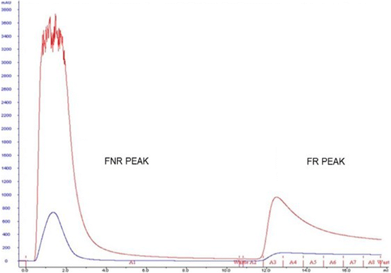
Methods: Serum samples were obtained from ten patients at the time of diagnosis (B-ALL group) and after induction therapy (AIT group). Sera from healthy children were used as controls (Control group). The samples were subjected to immunodepletion, affinity chromatography with α-d-galactose-binding lectin (from Artocarpus incisa seeds) immobilized on a Sepharose(TM) 4B gel, concentration, and digestion for subsequent analysis with nano-UPLC tandem nano-ESI-MS(E). The program Expression (E) was used to quantify differences in protein expression between groups.
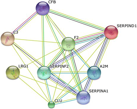
Results: A total of 96 proteins were identified. Leucine-rich alpha-2-glycoprotein 1 (LRG1), Clusterin (CLU), thrombin (F2), heparin cofactor II (SERPIND1), alpha-2-macroglobulin (A2M), alpha-2-antiplasmin (SERPINF2), Alpha-1 antitrypsin (SERPINA1), Complement factor B (CFB) and Complement C3 (C3) were identified as candidate biomarkers for early diagnosis of B-ALL, as they were upregulated in the B-ALL group relative to the control and AIT groups. Expression levels of the candidate biomarkers did not differ significantly between the AIT and control groups, providing further evidence that the candidate biomarkers are present only in the disease state, as all patients achieved complete remission after treatment.
Conclusion: A panel of protein biomarker candidates has been developed for pre-diagnosis of B-ALL and also provided information that would indicate a favorable response to treatment after induction therapy.
Keywords: Acute lymphoblastic leukemia; Biomarker; Frutalin; Lectin; Mass spectrometry.
Figures


Similar articles
-
Crystallization and preliminary X-ray diffraction studies of frutalin, an α-D-galactose-specific lectin from Artocarpus incisa seeds.Acta Crystallogr F Struct Biol Commun. 2015 Oct;71(Pt 10):1282-5. doi: 10.1107/S2053230X15015186. Epub 2015 Sep 23. Acta Crystallogr F Struct Biol Commun. 2015. PMID: 26457519 Free PMC article.
-
Identification of putative serum glycoprotein biomarkers for human lung adenocarcinoma by multilectin affinity chromatography and LC-MS/MS.Proteomics. 2007 Dec;7(23):4292-302. doi: 10.1002/pmic.200700433. Proteomics. 2007. PMID: 17963278
-
Identification and validation of serum autoantibodies in children with B-cell acute lymphoblastic leukemia by serological proteome analysis.Proteome Sci. 2022 Feb 2;20(1):3. doi: 10.1186/s12953-021-00184-w. Proteome Sci. 2022. PMID: 35109855 Free PMC article.
-
Serum Glycoprotein Biomarker Discovery and Qualification Pipeline Reveals Novel Diagnostic Biomarker Candidates for Esophageal Adenocarcinoma.Mol Cell Proteomics. 2015 Nov;14(11):3023-39. doi: 10.1074/mcp.M115.050922. Epub 2015 Sep 24. Mol Cell Proteomics. 2015. PMID: 26404905 Free PMC article.
-
Leucine-Rich Alpha-2-Glycoprotein as a non-invasive biomarker for pediatric acute appendicitis: a systematic review and meta-analysis.Eur J Pediatr. 2023 Jul;182(7):3033-3044. doi: 10.1007/s00431-023-04978-2. Epub 2023 May 6. Eur J Pediatr. 2023. PMID: 37148275 Free PMC article.
Cited by
-
Clinical Proteomics of Biofluids in Haematological Malignancies.Int J Mol Sci. 2021 Jul 27;22(15):8021. doi: 10.3390/ijms22158021. Int J Mol Sci. 2021. PMID: 34360786 Free PMC article. Review.
-
LRG1: an emerging player in disease pathogenesis.J Biomed Sci. 2022 Jan 21;29(1):6. doi: 10.1186/s12929-022-00790-6. J Biomed Sci. 2022. PMID: 35062948 Free PMC article. Review.
-
Proteomic analysis of cerebrospinal fluid in pediatric acute lymphoblastic leukemia patients: a pilot study.Onco Targets Ther. 2019 May 17;12:3859-3868. doi: 10.2147/OTT.S193616. eCollection 2019. Onco Targets Ther. 2019. PMID: 31190885 Free PMC article.
-
Circulating Biomarkers Associated with the Diagnosis and Prognosis of B-Cell Progenitor Acute Lymphoblastic Leukemia.Cancers (Basel). 2023 Aug 20;15(16):4186. doi: 10.3390/cancers15164186. Cancers (Basel). 2023. PMID: 37627214 Free PMC article. Review.
-
Dysregulation of Key Proteins Associated with Sperm Motility and Fertility Potential in Cancer Patients.Int J Mol Sci. 2020 Sep 15;21(18):6754. doi: 10.3390/ijms21186754. Int J Mol Sci. 2020. PMID: 32942548 Free PMC article.
References
-
- Scheurer ME, Bondy ML, Gurney JG. Epidemiology of Childhood Cancer. In: Pizzo PA, Poplack DG, editors. Principles and practice of pediatric oncology. 6. Philadelphia: Lippincott Williams and Wilkens; 2011. pp. 2–16.
-
- Michalowski MB, Lorea CF, Rech A, Santiago P, Lorenzoni M, Taniguchi A, et al. Diagnóstico precoce em oncologia pediátrica: uma urgência médica. Bol Cient Pediatr. 2012;01:13–8.
LinkOut - more resources
Full Text Sources
Other Literature Sources
Research Materials
Miscellaneous

