. 2015 Dec 11;1(11):e1500905.
doi: 10.1126/sciadv.1500905. eCollection 2015 Dec.

Engineering a nanopore with co-chaperonin function

Affiliations

Engineering a nanopore with co-chaperonin function

Ching-Wen Ho et al. Sci Adv. .

Abstract

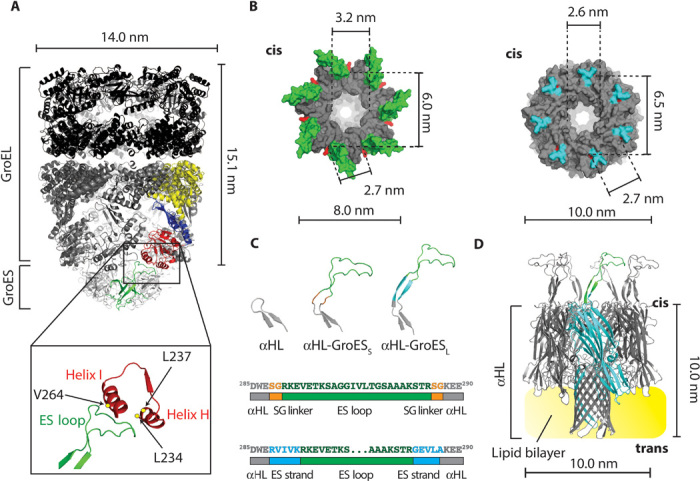
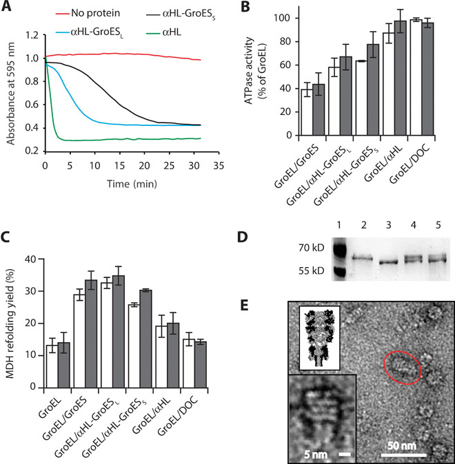
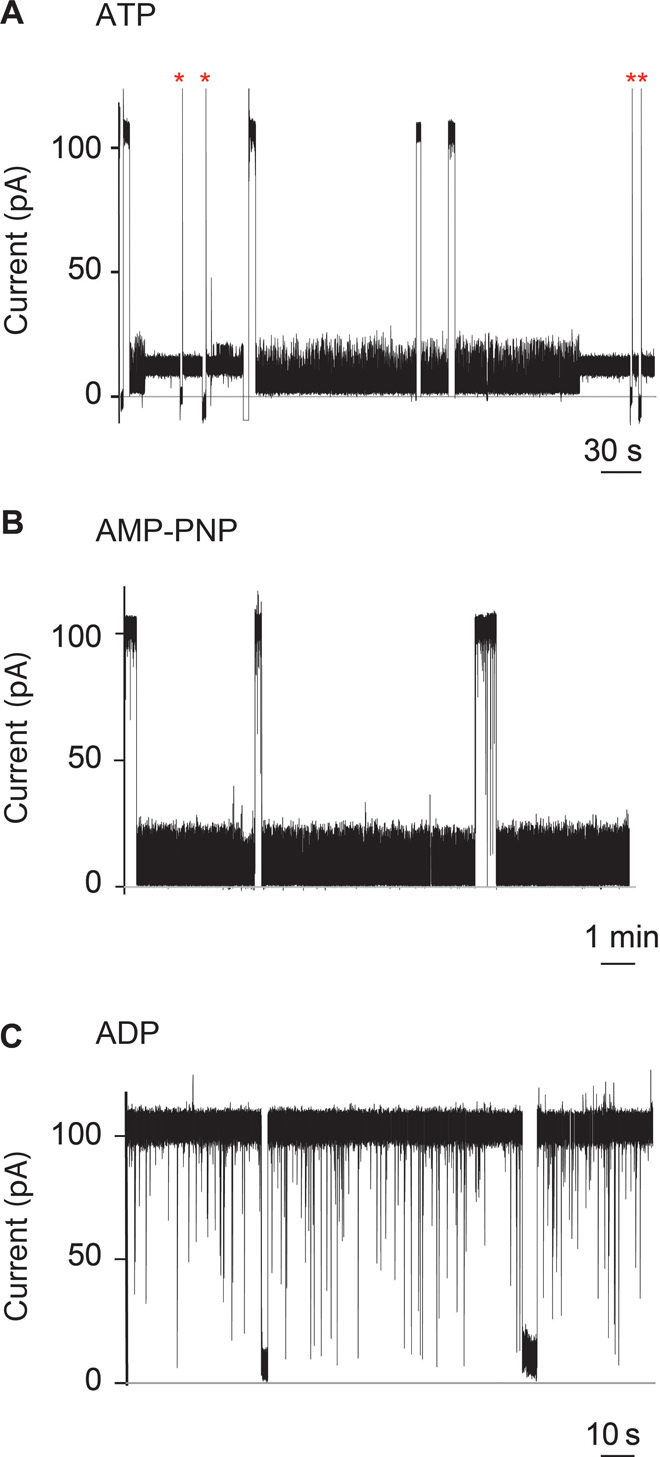
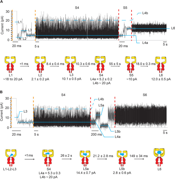
The emergence of an enzymatic function can reveal functional insights and allows the engineering of biological systems with enhanced properties. We engineered an alpha hemolysin nanopore to function as GroES, a protein that, in complex with GroEL, forms a two-stroke protein-folding nanomachine. The transmembrane co-chaperonin was prepared by recombination of GroES functional elements with the nanopore, suggesting that emergent functions in molecular machines can be added bottom-up by incorporating modular elements into preexisting protein scaffolds. The binding of a single-ring version of GroEL to individual GroES nanopores prompted large changes to the unitary nanopore current, most likely reflecting the allosteric transitions of the chaperonin apical domains. One of the GroEL-induced current levels showed fast fluctuations (<1 ms), a characteristic that might be instrumental for efficient substrate encapsulation or folding. In the presence of unfolded proteins, the pattern of current transitions changed, suggesting a possible mechanism in which the free energy of adenosine triphosphate binding and hydrolysis is expended only when substrate proteins are occupied.

Keywords: GroEL; GroES; Protein folding; loop grafting; nanomachine; single-molecule.


Figures

Fig. 1
Fig. 1. A GroEL:GroES-nanopore machine.
(A) Ribbon representation of the ADP:GroEL:GroES complex (PDB: 1AON). Single-GroES and single-GroEL subunits are colored: apical domain, red; equatorial domain, yellow; intermediate domain, blue; GroES subunit, green. The expansion shows the interaction between the ES loop and the H and I helices of the GroEL apical domain. The amino acids in GroEL that are mainly involved in the interaction with GroES (L234, L237, and V264) are shown as yellow dots. (B) Surface representation of the top view of the cis side of GroES (left, PDB: 1AON) and αHL (right, PDB: 7AHL). The ES loops are green and the αHL loops are cyan. The lines indicate the diameter of the cis central apertures and the α-carbon distance of the first residue (red) of the ES loops (K13) or the αHL loops (W286) between adjacent and opposite subunits. (C) Top, β-hairpin loops in αHL (left), αHL-GroESS (center), and αHL-GroESL (right). Bottom, scheme describing ES loop insertions (green) into the αHL sequence (gray) for αHL-GroESS (top) and αHL-GroESL (bottom) constructs. The SG linker is depicted in orange and the ES strand (10 additional GroES residues that form a β strand in GroES) is shown in blue. (D) Ribbon representation of αHL-GroESL (gray) prepared by homology modeling from the αHL and GroES structures. An αHL subunit is shown in cyan with one ES loop in green, and the lipid bilayer is shown in yellow.
Fig. 2
Fig. 2. Characterization of αHL-GroES chimera proteins.
(A) Typical traces for the hemolytic assay showing the pore-forming activity of nanopore proteins. Each line shows the reduction in absorbance for monomeric nanopore proteins: αHL is in green, αHL-GroESL is in blue, and αHL-GroESS is in black. The red line shows a control experiment where no protein was used. Proteins are added to a 0.0625 μM final concentration and were incubated with a solution of diluted rabbit red blood cells in 10 mM Mops and 150 mM NaCl (pH 7.4) containing bovine serum albumin (1 mg/ml). The pore-forming activity of the αHL proteins is shown by the decrease in the absorbance at 595 nm due to the lysis of the rabbit red blood cells. (B) ATPase activity of GroEL (50 nM) in the presence of GroES proteins (200 nM) and/or the surfactant used to solubilize the nanopores [deoxycholate (DOC), 0.125 mM] in 50 mM KCl (white bars) or 1 M KCl (gray bars). The ATPase reaction was started by adding ATP to the reaction buffer containing GroES and GroEL proteins. The values are shown in table S2. (C) Refolding assay assisted by GroES proteins in 50 mM KCl (white bars) or 1 M KCl (gray bars). Unfolded MDH (2 μM) was preincubated with GroEL (50 nM) in the refolding buffer (no ATP) before the addition of ATP (2 mM) and co-chaperonin proteins (200 nM). Control experiments with DOC and αHL indicated that the GroEL-assisted refolding is mediated by the grafted loops in αHL-GroESL. Refolding yields were normalized by the spontaneous refolding of MDH. The values are shown in table S3. (D) Proteinase K protection assay. GroEL (0.2 μM), preincubated with GroES or αHL-GroESL (1 μM) in lanes 4 and 5, respectively, was treated with proteinase K (25 μg/ml) in the presence of ATP (1 mM) for 20 min before loading into polyacrylamide SDS gels. Lane 1, protein ladder. Lane 2, undigested GroEL (58 kD). Lane 3, GroEL after incubation with proteinase K (~56 kD). Lane 4, GroEL digestion protected by GroES. Lane 5, GroEL digestion protected by αHL-GroESL. (E) Negative-stained EM image of GroEL-398 bound to αHL-GroESL formed by preincubating GroEL-398 (0.5 μM) with αHL-GroESL (1 μM) for 20 min before applying a 100-fold dilution to negatively stained EM grids. The insets show the magnification of the circled complex (bottom) and a scaled surface representation of the αHL-GroESL:GroEL complex (top). Experiments and dilutions in (B) and (C) were carried out in 50 mM tris-HCl (pH 7.5), 50 mM KCl, 1 mM ATP, and 5 mM MgCl2. Errors are quoted as SD.
Fig. 3
Fig. 3. Interaction of SR1 with membrane-bound αHL-GroESL monitored by single-molecule recordings.
(A to C) Current blockades provoked by SR1 to αHL-GroESL in the presence of 1 mM ATP (A), the nonhydrolyzable ATP analog AMP-PNP (B), or adenosine diphosphate (ADP) (C) added to the cis chamber at +100 mV. The current blockades are due to the interaction of the chaperonin with individual engineered co-chaperonin nanopores. (A) Fifty percent of the blockades in the presence of ATP were transient (dwell time, 55 ± 5 s; main IB value, 5.2 ± 0.2 pA), showing fast current fluctuations (see the text and fig. S5). The remaining current blockades switched to a quieter and permanent current level (IB value, 12.5 ± 0.5 pA). The open pore current could only be obtained by multiple ramping of the potential to +100/−100 mV (red asterisks). (B) AMP-PNP–induced current blockades showed the same current level as ATP-induced blockades but were always transient (average dwell time, 157 ± 64 s; main IB value, 5.2 ± 0.1 pA) (see the text). (C) In the presence of ADP, the current blockades were transient, showing a dwell time faster than the resolution of our current recordings (dwell time, <1 ms). Occasionally longer current blockades were observed, which showed an IB value of 3.0 ± 1.4 pA (fig. S6c). The electrical recordings were carried out in 1 M KCl, 50 mM tris-HCl (pH 7.5), and 5 mM MgCl2 at 23°C and +100 mV by applying a 10-kHz low-pass Bessel filter with a 50-kHz sampling rate. An additional digital Gaussian filter at 2 kHz was applied to current traces.
Fig. 4
Fig. 4. Intermediates of SR1 protein-folding cycle.
Typical SR1 current blockades. The current traces between the vertical dotted orange and red lines are expansions of the main current traces showing the fast current transitions that characterize the L1, L2, L3, and S5 states. The cartoon depictions below the current traces show the kinetic succession between SR1 intermediates. SR1 is in yellow, αHL-GroESL is in red, and the substrate protein is in blue. (A) αHL-GroESL current blockade for the ternary SR1:ATP:αHL-GroESL complex. L1 (dwell time, <1 ms) was observed only in a few blockades (6%). Ninety-two percent of blockades started with either L2 (77%) or L3 (15%) current levels. The transition from S4 to L6 was fast (10.3 ± 0.6 ms; fig. S5) and characterized by current fluctuations showing an average value at ~10 pA. An expansion of the S4 current state is shown in fig. S5. (B) αHL-GroESL current blockade for the quaternary SR1:ATP:DHFR:αHL-GroESL complex. In 91% of blockades, L1, L2, and L3 current levels could not be sampled, whereas the transition between S4 and L6 showed two well-defined current levels, indicating that the unfolded protein modified the interaction of SR1 with αHL-GroESL (see the text). Dihydrofolate reductase (DHFR) was urea unfolded and incubated with SR1 before addition to the cis chamber. Traces were recorded in 1 M KCl and 50 mM tris-HCl (pH 7.5) at 23°C in the presence of 1 mM ATP and 5 mM MgCl2 by applying a 10-kHz low-pass Bessel filter and using a 20-μs (50-kHz) sampling rate. An additional postacquisition Gaussian filter at 2 kHz was then applied to the current traces.

Similar articles

Cited by

References

    1. Maglia G., Henricus M., Wyss R., Li Q., Cheley S., Bayley H., DNA strands from denatured duplexes are translocated through engineered protein nanopores at alkaline pH. Nano Lett. 9, 3831–3836 (2009). - PubMed
    1. Japrung D., Henricus M., Li Q., Maglia G., Bayley H., Urea facilitates the translocation of single-stranded DNA and RNA through the α-hemolysin nanopore. Biophys. J. 98, 1856–1863 (2010). - PMC - PubMed
    1. Biesemans A., Soskine M., Maglia G., A protein rotaxane controls the translocation of proteins across a ClyA nanopore. Nano Lett. 15, 6076–6081 (2015). - PMC - PubMed
    1. Sánchez-Quesada J., Saghatelian A., Cheley S., Bayley H., Ghadiri M. R., Single DNA rotaxanes of a transmembrane pore protein. Angew. Chem. Int. Ed. 43, 3063–3067 (2004). - PMC - PubMed
    1. Bezrukov S. M., Vodyanoy I., Parsegian V. A., Counting polymers moving through a single ion channel. Nature 370, 279–281 (1994). - PubMed