

Quantification of cells expressing markers of proliferation and apoptosis in chronic tonsilitis
- PMID: 26824215
- PMCID: PMC4731890
Quantification of cells expressing markers of proliferation and apoptosis in chronic tonsilitis
Abstract
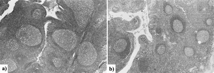
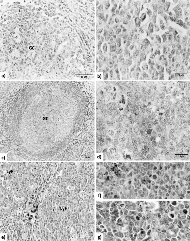
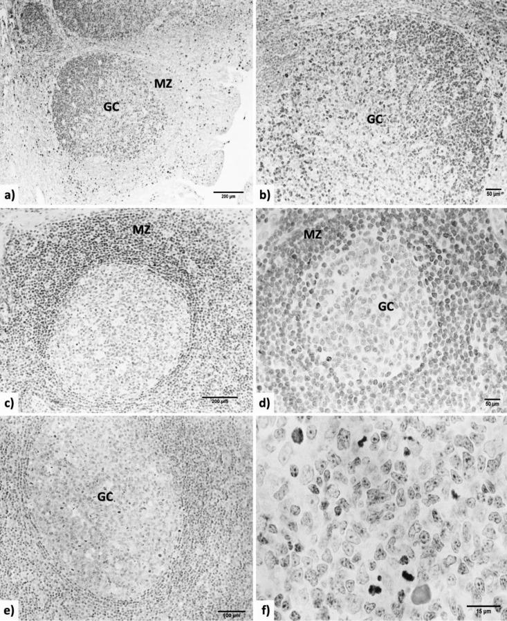
During chronic tonsillitis, the relationship between proliferation and apoptosis of lymphocytes in tonsillar follicles can be disturbed, which gives rise to attenuation of tonsil immunocompetence and diminishing its contribution in systemic immunity. In this study, we have quantified the cells expressing the markers of proliferation and apoptosis in the follicles of the palatine tonsil. Six tonsils from patients aged 10-29 years with hypertrophic tonsillitis and five tonsils from patients aged 18-22 years with recurrent tonsillitis were studied. The sections of paraffin blocks of tonsillar tissue were stained by the immunohistochemical LSAB/HRP method with the utilisation of antibodies for: Ki-67 antigen-cell marker of proliferation; Bcl-2 and survivin anti-apoptotic factors and Fas/CD95, caspase-3 and Bax pro-apoptotic factors. The size of lymphoid follicles, i.e. mean follicle area and number of lymphoid follicle immunopositive cells per mm2 of a slice area, i.e. numerical areal density were determined by the quantitative image analysis. The localisation of Ki-67, Bcl-2, survivin, Fas/CD95, caspase-3 and Bax- immunopositive cells inside the palatine tonsil was similar in both types of tonsillitis. The number of Ki-67 immunopositive cells was significantly (p < 0.01) larger in the tonsils with hypertrophic tonsillitis (14681.4 ± 1460.5) in comparison to those with recurrent tonsillitis (12491.4 ± 2321.6), although the number of survivin and caspase-3 immunopositive cells was significantly (p < 0.05) larger in recurrent tonsillitis (survivin, 406.9 ± 98.4; caspase-3, 350.4 ± 119.4) when compared to those with hypertrophic tonsillitis (survivin, 117.4 ± 14.5; caspase-3, 210 ± 24). Our results show that the rate of the proliferation and apoptosis of follicular lymphocytes is different in various types of tonsillitis. This suggests that the immunological potential of the palatine tonsil varies in patients with hypertrophic and recurrent tonsillitis, which in practice poses a dilemma over the choice of conservative or surgical treatment.
Durante una tonsillite cronica, il rapporto tra proliferazione e apoptosi dei linfociti nei follicoli tonsillari può essere alterato: ciò spiega l'attenuazione dell'immunocompetenza tonsillare e la riduzione del suo contributo all' immunità sistemica. In questo studio abbiamo quantificato le cellule che esprimono i marker di proliferazione e apoptosi nei follicoli delle tonsille palatine. Sono state studiate sei tonsille da pazienti di età compresa tra 10 e 29 anni con tonsillite ipertrofica e cinque tonsille da pazienti di età compresa tra 18 e 22 anni con tonsillite ricorrente. Le sezioni di tessuto tonsillare incluso in paraffina sono state colorate con il metodo immunoistochimico di LSAB/HRP attraverso l'applicazione di anticorpi per: l'antigene ki-67, marcatore cellulare di proliferazione; Bcl-2 e survivina, fattori antiapoptotici; Fas/CD95, caspasi 3 e Bax, fattori proapoptotici. La dimensione dei follicoli linfatici, ossia l'area del follicolo e il numero delle cellule immunopositive del follicolo per mm2, ossia la densità numerica dell'area, sono state determinate attraverso una analisi quantitativa dell'immagine. La localizzazione delle cellule immunopositive a ki-67, Bcl-2, survivina, Fas/CD95, caspasi 3 e Bax all'interno della tonsilla palatina è stata simile nei due tipi di tonsillite. Il numero delle cellule immunopositive per Ki-67 è stato significativamente (p < 0,01) maggiore nelle tonsille con tonsillite ipertrofica (14681,4 ± 1460,5) rispetto a quelle con tonsillite ricorrente (12491,4 ± 2321,6), sebbene il numero di cellule immunopositive per survivina e caspasi 3 fosse significativamente (p < 0,05) maggiore nelle tonsilliti ricorrenti (survivina, 406,9 ± 98,4; caspasi-3, 350,4 ± 119,4) rispetto alle tonsille con tonsilliti ipertrofiche (survivina, 117,4 ± 14,5; caspasi-3, 210 ± 24). I nostri risultati mostrano che il tasso di proliferazione e apoptosi dei linfociti follicolari è diverso nei vari tipi di tonsillite. Questo indica che il potenziale immunologico della tonsilla palatina varia nei pazienti con tonsillite ipertrofica rispetto a quelli con tonsillite ricorrente: ciò, nella pratica, pone un dilemma in merito alla scelta del trattamento conservativo o chirurgico.
Keywords: Apoptosis; Cell proliferation; Human palatine tonsil; Lymphoid follicle; Quantification.
Figures
Similar articles
-
Lack of lymphoid cell apoptosis in the pathogenesis of tonsillar hypertrophy as compared to recurrent tonsillitis.Eur J Pediatr. 1999 Jun;158(6):469-73. doi: 10.1007/s004310051122. Eur J Pediatr. 1999. PMID: 10378394
-
Apoptosis in chronic tonsillitis and tonsillar hypertrophy.Int J Pediatr Otorhinolaryngol. 2015 Feb;79(2):191-5. doi: 10.1016/j.ijporl.2014.12.005. Epub 2014 Dec 15. Int J Pediatr Otorhinolaryngol. 2015. PMID: 25555639
-
Comparison of histology between recurrent tonsillitis and tonsillar hypertrophy.Clin Otolaryngol Allied Sci. 2003 Jun;28(3):235-9. doi: 10.1046/j.1365-2273.2003.00697.x. Clin Otolaryngol Allied Sci. 2003. PMID: 12755763
-
Peritonsillar abscess: clinical aspects of microbiology, risk factors, and the association with parapharyngeal abscess.Dan Med J. 2017 Mar;64(3):B5333. Dan Med J. 2017. PMID: 28260599 Review.
-
Is tonsillectomy mandatory for asymmetric tonsils in children? A review of our diagnostic tonsillectomy practice and the literature.Int J Pediatr Otorhinolaryngol. 2018 Jul;110:57-60. doi: 10.1016/j.ijporl.2018.04.027. Epub 2018 May 1. Int J Pediatr Otorhinolaryngol. 2018. PMID: 29859588 Review.
Cited by
-
Role of Apoptosis in Wound Healing and Apoptosis Alterations in Microgravity.Front Bioeng Biotechnol. 2021 Jun 17;9:679650. doi: 10.3389/fbioe.2021.679650. eCollection 2021. Front Bioeng Biotechnol. 2021. PMID: 34222218 Free PMC article. Review.
-
Recent advances in molecular mechanisms of skin wound healing and its treatments.Front Immunol. 2024 May 21;15:1395479. doi: 10.3389/fimmu.2024.1395479. eCollection 2024. Front Immunol. 2024. PMID: 38835782 Free PMC article. Review.
-
Death of tonsillar B cells by NETosis.Cell Death Discov. 2023 Mar 30;9(1):108. doi: 10.1038/s41420-023-01402-4. Cell Death Discov. 2023. PMID: 36997529 Free PMC article.
References
-
- Perry M, Whyte A. Immunology of the tonsils. Immunol Today. 1998;19:414–421. - PubMed
-
- Nave H, Gebert A, Pabst R. Morphology and immunology of the human palatine tonsil. Anat Embryol (Berl) 2001;204:367–373. - PubMed
-
- Brandtzaeg P. Immunology of tonsils and adenoids: everything the ENT surgeon needs to know. Int J Pediatr Otorhinolaryngol. 2003;67:s69–s76. - PubMed
Publication types
MeSH terms
Substances
LinkOut - more resources
Full Text Sources
Medical
Research Materials
Miscellaneous