. 2016 Feb 23;7(8):9118-34.
doi: 10.18632/oncotarget.7001.

MIIP accelerates epidermal growth factor receptor protein turnover and attenuates proliferation in non-small cell lung cancer

Affiliations

MIIP accelerates epidermal growth factor receptor protein turnover and attenuates proliferation in non-small cell lung cancer

Jing Wen et al. Oncotarget. .

Abstract

The migration and invasion inhibitory protein (MIIP) has been discovered recently to have inhibitory functions in cell proliferation and migration. Overexpression of MIIP reduced the intracellular steady-state level of epidermal growth factor receptor (EGFR) protein in lung cancer cells with no effect on EGFR mRNA expression compared to that in the control cells. This MIIP-promoted EGFR protein degradation was reversed by proteasome and lysosome inhibitors, suggesting the involvement of both proteasomal and lysosomal pathways in this degradation. This finding was further validated by pulse-chase experiments using 35S-methionine metabolic labeling. We found that MIIP accelerates EGFR protein turnover via proteasomal degradation in the endoplasmic reticulum and then via the lysosomal pathway after its entry into endocytic trafficking. MIIP-stimulated downregulation of EGFR inhibits downstream activation of Ras and blocks the MEK signal transduction pathway, resulting in inhibition of cell proliferation. The negative correlation between MIIP and EGFR protein expression was validated in lung adenocarcinoma samples. Furthermore, the higher MIIP protein expression predicts a better overall survival of Stage IA-IIIA lung adenocarcinoma patients who underwent radical surgery. These findings reveal a new mechanism by which MIIP inhibits cell proliferation.

Keywords: epidermal growth factor receptor; migration and invasion inhibitory protein; non-small cell lung cancer; protein degradation.


Conflict of interest statement

CONFLICTS OF INTEREST

The authors declare no conflict of interest.

Figures
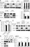
Figure 1
Figure 1. Inverse patterns of MIIP and EGFR protein expression in human lung cancer cell lines
A. Western blotting analysis of steady-state EGFR protein levels in H1299, A549, and H322 cells transfected with MIIP-HA plasmid or with shMIIP. All error bars show standard error for triplicate experiments. *, P < 0.05; ***, P < 0.001; NS, not significant by Student t-test. B. Real-time RT-PCR analysis of EGFR mRNA levels in MIIP-HA−overexpressing or MIIP-knockdown cells. All error bars show standard error for triplicate experiments. NS, not significant by Student t-test. C. Western blotting analysis of steady-state EGFR protein levels in MIIP-HA−overexpressing or control H1299 cells with 10 μM lactacystin or 100 μM chloroquine treatment for 2 h. Numbers below the western blotting bands represents the quantification of gel densitometry. D. Immunoprecipitation (IP) and immunoblotting (IB) assay of ubiquitinated EGFR in MIIP-HA−overexpressing or control H1299 cells with blank protein G beads as negative control. Proteins bound to anti-EGFR-conjugated protein G beads were collected and subjected to IB analysis. INPUT, immunoblot of steady levels of EGFR, ubiquitin, MIIP-HA, and β-actin in cell lysate (10% of the same cell lysate samples used for immunoprecipitation). The intensities of the bands were quantified with densitometry and normalized with that of β-actin loading control. Values are presented in bar graphs as percentage relative to control (100%). All error bars show standard error for triplicate experiments. **, P < 0.01; ***, P < 0.001; NS, not significant by Student t-test.
Figure 2
Figure 2. MIIP accelerates EGFR protein turnover
Intracellular stability of endogenous EGFR protein level was measured by protein turnover assay in H1299 cells stably transfected with MIIP and in control cells. Cells were radiolabeled with 35S-methionine in a pulse-chase experiment, and collected at indicated chase time points. Clarified cell lysates were used for immunoprecipitation of endogenous EGFR. Immunoprecipitated proteins were resolved on SDS-PAGE and detected by fluorography. A. Increased turnover rate of endogenous EGFR protein was shown in cells stably transfected with MIIP. B. Graphic representation of EGFR protein turnover is based on quantification of gel densitometry. All error bars show standard error for triplicate experiments. **, P < 0.01 by repeated measures ANOVA.
Figure 3
Figure 3. MIIP promotes degradation of newly synthesized EGFR by the proteasome pathway
H1299 cells stably transfected with MIIP and control cells were radiolabeled with 35S-methionine for indicated time in a pulse experiment without drug treatment (Ctrl; A) or with 10 μM lactacystin (Lac; B) or 5 μM brefeldin A (BFA; C) treatment. Clarified cell lysates were used for immunoprecipitation of endogenous EGFR. Graphic representation of EGFR protein turnover is based on quantification of gel densitometry from triplicate experiments. A. Turnover of newly synthesized endogenous EGFR in MIIP-HA−overexpressing or control H1299 cells. ***, P < 0.001 by repeated measures ANOVA. B. Turnover of newly synthesized endogenous EGFR in MIIP-HA−overexpressing and control H1299 cells with lactacystin treatment. NS, not significant by repeated measures ANOVA. C. Turnover of newly-synthesized endogenous EGFR in MIIP-HA−overexpressing or control H1299 cells with brefeldin A treatment. **, P < 0.01 by repeated measures ANOVA. D. Reciprocal co-immunoprecipitation (IP) assay of endogenous EGFR and BIP in MIIP-HA−overexpressing or control H1299 cells with blank protein G beads as negative control. Proteins bound to protein G beads were collected and subjected to SDS-PAGE/western blotting (IB) analysis. INPUT, immunoblot of steady levels of EGFR, BIP, MIIP-HA, and β-actin in cell lysate (10% of the same cell lysate samples used for immunoprecipitation). E. The intensities of the bands in D were quantified with densitometry and normalized with that of β-actin loading control. Values are presented in bar graphs as percentage relative to control (100%). All error bars show standard error for triplicate experiments. *, P < 0.05; NS, not significant by Student t-test.
Figure 4
Figure 4. MIIP accelerates lysosomal degradation of mature EGFR
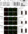
A. H1299 cells stably transfected with MIIP-HA and control plasmid were radiolabeled with 35S-methionine for 40 min and chased for indicated time without (Ctrl) or with 100 μM chloroquine (CQ). Graphic representation of EGFR protein turnover is based on quantification of gel densitometry from triplicate experiments. NS, not significant; **, P < 0.01 by Student t-test. B. Western blotting determined expression of steady-state HDAC6 protein in freshly MIIP-HA-transfected and control H1299 cells. C. H1299 cells stably transfected with MIIP-HA and control plasmid were treated with 10ng/ml EGF for 5min, chased for the indicated time, and then subjected to immunofluorescence staining with anti-EGFR (red) and anti-EEA1 (green). Quantification of colocalization between EGFR and EEA1 signals is shown in the histograms. NS, not significant by Student t-test. (Scale bar: 40μM) D. H1299 cells stably transfected with MIIP-HA and control plasmid were treated as described in B and immunofluorescent stained with anti-EGFR (red) and anti-LAMP1 (green). Quantification of colocalization between EGFR and LAMP1 signals is shown in the histograms. NS, not significant; **, P < 0.01 by Student t-test. (Scale bar: 40μM) E. H1299 cells stably transfected with MIIP-HA and control plasmid were treated as described in B and immunofluorescent stained with anti-EGFR (red) and anti-Rab11 (green). Quantification of colocalization between EGFR and Rab11 signals is shown in the histograms. NS, not significant; **, P < 0.01 by Student t-test. (Scale bar: 40μM)
Figure 5
Figure 5. Functional analysis of the effects of MIIP expression on EGFR's EGF-binding activity and activation of EGFR and downstream signaling and cell proliferation
A. EGF-binding activity was determined in MIIP-HA−overexpressing and control H1299 cells. EGFR bound to the EGF-conjugated beads was pulled down and subjected to immunoblotting with anti-EGFR antibody. Ten percent of each original total cell lysate was used as input quantification, using β-actin as loading control. All error bars show standard error for triplicate experiments. *, P < 0.05; **, P < 0.01; ***, P < 0.001; NS, not significant by Student t-test. B. Activation of Ras and the MEK/MAPK pathway in MIIP-HA−overexpressing or MIIP-knockdown H1299 cells. Numbers below the western blotting bands represents the quantification of gel densitometry. C. Cell proliferation was determined in MIIP-HA−overexpressing or MIIP-knockdown H1299 and A549 cells. Single-cell suspensions prepared from freshly selected cells positive for MIIP overexpression or MIIP knockdown were seeded in 6-well plates, and counted at indicated time points in triplicate. *, P < 0.05; **, P < 0.01; NS, not significant by repeated measures ANOVA.
Figure 6
Figure 6. MIIP protein expression correlates negatively with EGFR protein expression in lung adenocarcinoma specimens and predicts patients' survivals
A. Representative immunohistochemical staining of a lung adenocarcinoma case with low MIIP and high EGFR expression. (Scale bar: 40μM) B. Representative immunohistochemical staining of a lung adenocarcinoma case with high MIIP and low EGFR expression. (Scale bar: 40μM) C. Correlation between MIIP and EGFR protein expression in 28 lung adenocarcinoma specimens (Spearman's rank correlation analysis, r = −0.473, P = 0.011). D. Kaplan-Meier survival analysis of 234 Stage IA to IIIA lung adenocarcinoma patients with different MIIP expression levels (P = 0.043 by log-rank test). E. MIIP accelerates EGFR protein turnover, which results in the inhibition of lung cancer cell proliferation. MIIP enhances the binding between newly-synthesized EGFR protein and BIP, leads to the degradation of the nascent EGFR through the proteasome pathway. In addition, MIIP could increase the trafficking of mature EGFR into late endosomes and subsequent degradation in lysosome and decrease EGFR recycling back to cellular membrane by directly decreasing HDAC6 expression, one of the downstream targets of MIIP. The downregulation of steady-state EGFR by MIIP results in less activation of EGFR and downstream Ras/MEK/ERK pathway, which finally inhibits lung cancer cell proliferation.

Similar articles

Cited by

References

    1. Torre LA, Bray F, Siegel RL, Ferlay J, Lortet-Tieulent J, Jemal A. Global cancer statistics, 2012. CA Cancer J Clin. 2015;65:87–108. - PubMed
    1. Chen W, Zheng R, Zeng H, Zhang S. The updated incidences and mortalities of major cancers in China, 2011. Chin J Cancer. 2015;34:53. - PMC - PubMed
    1. Wykosky J, Fenton T, Furnari F, Cavenee WK. Therapeutic targeting of epidermal growth factor receptor in human cancer: successes and limitations. Chin J Cancer. 2011;30:5–12. - PMC - PubMed
    1. Xu N, Fang W, Mu L, Tang Y, Gao L, Ren S, Cao D, Zhou L, Zhang A, Liu D, Zhou C, Wong KK, Yu L, et al. Overexpression of wildtype EGFR is tumorigenic and denotes a therapeutic target in non-small cell lung cancer. Oncotarget. 2015 doi: 10.18632/oncotarget.6461. - DOI - PMC - PubMed
    1. Hirsch FR, Varella-Garcia M, Cappuzzo F. Predictive value of EGFR and HER2 overexpression in advanced non-small-cell lung cancer. Oncogene. 2009;28(Suppl 1):S32–37. - PubMed

Publication types

MeSH terms