WISP-1 promotes VEGF-C-dependent lymphangiogenesis by inhibiting miR-300 in human oral squamous cell carcinoma cells
- PMID: 26824419
- PMCID: PMC4891098
- DOI: 10.18632/oncotarget.7014
WISP-1 promotes VEGF-C-dependent lymphangiogenesis by inhibiting miR-300 in human oral squamous cell carcinoma cells
Abstract
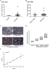
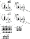
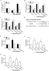
Oral squamous cell carcinoma (OSCC), which accounts for nearly 90% of head and neck cancers, is characterized by a poor prognosis and a low survival rate. Vascular endothelial growth factor-C (VEGF-C) has been implicated in lymphangiogenesis and is correlated with cancer metastasis. WNT1-inducible signaling pathway protein-1 (WISP)-1/CCN4 is an extracellular matrix-related protein that belongs to the CCN family and stimulates many biological functions. Our previous studies showed that WISP-1 plays an important role in OSCC migration and angiogenesis. However, the effect of WISP-1 on VEGF-C regulation and lymphangiogenesis in OSCC is poorly understood. Here, we showed a correlation between WISP-1 and VEGF-C in tissue specimens from patients with OSCC. To examine the lymphangiogenic effect of WISP-1, we used human lymphatic endothelial cells (LECs) to mimic lymphatic vessel formation. The results showed that conditioned media from WISP-1-treated OSCC cells promoted tube formation and cell migration in LECs. We also found that WISP-1-induced VEGF-C is mediated via the integrin αvβ3/integrin-linked kinase (ILK)/Akt signaling pathway. In addition, the expression of microRNA-300 (miR-300) was inhibited by WISP-1 via the integrin αvβ3/ILK/Akt cascade. Collectively, these results reveal the detailed mechanism by which WISP-1 promotes lymphangiogenesis via upregulation of VEGF-C expression in OSCC. Therefore, WISP-1 could serve as therapeutic target to prevent metastasis and lymphangiogenesis in OSCC.
Keywords: OSCC; VEGF-C; WISP-1; lymphangiogenesis; miR-300.
Conflict of interest statement
All authors have no financial or personal relationships with other people or organizations that could inappropriately influence our work.
Figures


Similar articles
-
WISP-1 a novel angiogenic regulator of the CCN family promotes oral squamous cell carcinoma angiogenesis through VEGF-A expression.Oncotarget. 2015 Feb 28;6(6):4239-52. doi: 10.18632/oncotarget.2978. Oncotarget. 2015. PMID: 25738362 Free PMC article.
-
Chemokine CCL4 Induces Vascular Endothelial Growth Factor C Expression and Lymphangiogenesis by miR-195-3p in Oral Squamous Cell Carcinoma.Front Immunol. 2018 Mar 2;9:412. doi: 10.3389/fimmu.2018.00412. eCollection 2018. Front Immunol. 2018. PMID: 29599774 Free PMC article.
-
Apoptosis signal-regulating kinase 1 is involved in WISP-1-promoted cell motility in human oral squamous cell carcinoma cells.PLoS One. 2013 Oct 21;8(10):e78022. doi: 10.1371/journal.pone.0078022. eCollection 2013. PLoS One. 2013. PMID: 24205072 Free PMC article.
-
The role of novel prognostic markers PROX1 and FOXC2 in carcinogenesis of oral squamous cell carcinoma.J Exp Ther Oncol. 2018 May;12(3):171-184. J Exp Ther Oncol. 2018. PMID: 29790306 Review.
-
Roles of the TGF-β⁻VEGF-C Pathway in Fibrosis-Related Lymphangiogenesis.Int J Mol Sci. 2018 Aug 23;19(9):2487. doi: 10.3390/ijms19092487. Int J Mol Sci. 2018. PMID: 30142879 Free PMC article. Review.
Cited by
-
The role of CCN4/WISP-1 in the cancerous phenotype.Cancer Manag Res. 2018 Aug 27;10:2893-2903. doi: 10.2147/CMAR.S133915. eCollection 2018. Cancer Manag Res. 2018. PMID: 30214284 Free PMC article. Review.
-
MicroRNA-433 inhibits oral squamous cell carcinoma cells by targeting FAK.Oncotarget. 2017 Oct 27;8(59):100227-100241. doi: 10.18632/oncotarget.22151. eCollection 2017 Nov 21. Oncotarget. 2017. Retraction in: Oncotarget. 2022 Sep 14;13:1033. doi: 10.18632/oncotarget.28270. PMID: 29245973 Free PMC article. Retracted.
-
The Roles of Non-Coding RNAs in Tumor-Associated Lymphangiogenesis.Cancers (Basel). 2020 Nov 6;12(11):3290. doi: 10.3390/cancers12113290. Cancers (Basel). 2020. PMID: 33172072 Free PMC article. Review.
-
WISP-1 Regulates Cardiac Fibrosis by Promoting Cardiac Fibroblasts' Activation and Collagen Processing.Cells. 2024 Jun 6;13(11):989. doi: 10.3390/cells13110989. Cells. 2024. PMID: 38891121 Free PMC article.
-
The Regulation of Lymph Node Pre-Metastatic Niche Formation in Head and Neck Squamous Cell Carcinoma.Front Oncol. 2022 Apr 27;12:852611. doi: 10.3389/fonc.2022.852611. eCollection 2022. Front Oncol. 2022. PMID: 35574333 Free PMC article. Review.
References
-
- Johnson NW, Jayasekara P, Amarasinghe AA. Squamous cell carcinoma and precursor lesions of the oral cavity: epidemiology and aetiology. Periodontol 2000. 2011;57:19–37. - PubMed
-
- Johnson NW, Warnakulasuriya S, Gupta PC, Dimba E, Chindia M, Otoh EC, Sankaranarayanan R, Califano J, Kowalski L. Global oral health inequalities in incidence and outcomes for oral cancer: causes and solutions. Adv Dent Res. 2011;23:237–246. - PubMed
-
- Kim SY, Nam SY, Choi SH, Cho KJ, Roh JL. Prognostic value of lymph node density in node-positive patients with oral squamous cell carcinoma. Ann Surg Oncol. 2011;18:2310–2317. - PubMed
-
- Eckert AW, Lautner MH, Schutze A, Bolte K, Bache M, Kappler M, Schubert J, Taubert H, Bilkenroth U. Co-expression of Hif1alpha and CAIX is associated with poor prognosis in oral squamous cell carcinoma patients. J Oral Pathol Med. 2010;39:313–317. - PubMed
-
- Ling N, Gu J, Lei Z, Li M, Zhao J, Zhang HT, Li X. microRNA-155 regulates cell proliferation and invasion by targeting FOXO3a in glioma. Oncol Rep. 2013;30:2111–8. - PubMed
Publication types
MeSH terms
Substances
LinkOut - more resources
Full Text Sources
Other Literature Sources
Medical
Research Materials

