. 2016 Jan 29;11(1):e0147736.
doi: 10.1371/journal.pone.0147736. eCollection 2016.

K+ Channel Inhibition Differentially Regulates Migration of Intestinal Epithelial Cells in Inflamed vs. Non-Inflamed Conditions in a PI3K/Akt-Mediated Manner

Affiliations

K+ Channel Inhibition Differentially Regulates Migration of Intestinal Epithelial Cells in Inflamed vs. Non-Inflamed Conditions in a PI3K/Akt-Mediated Manner

Sebastian Zundler et al. PLoS One. .

Abstract

Background: Potassium channels have been shown to determine wound healing in different tissues, but their role in intestinal epithelial restitution--the rapid closure of superficial wounds by intestinal epithelial cells (IEC)--remains unclear.

Methods: In this study, the regulation of IEC migration by potassium channel modulation was explored with and without additional epidermal growth factor (EGF) under baseline and interferon-γ (IFN-γ)-pretreated conditions in scratch assays and Boyden chamber assays using the intestinal epithelial cell lines IEC-18 and HT-29. To identify possibly involved subcellular pathways, Western Blot (WB)-analysis of ERK and Akt phosphorylation was conducted and PI3K and ERK inhibitors were used in scratch assays. Furthermore, mRNA-levels of the potassium channel KCNN4 were determined in IEC from patients suffering from inflammatory bowel diseases (IBD).

Results: Inhibition of Ca(2+)-dependent potassium channels significantly increased intestinal epithelial restitution, which could not be further promoted by additional EGF. In contrast, inhibition of KCNN4 after pretreatment with IFN-γ led to decreased or unaffected migration. This effect was abolished by EGF. Changes in Akt, but not in ERK phosphorylation strongly correlated with these findings and PI3K but not ERK inhibition abrogated the effect of KCNN4 inhibition. Levels of KCNN4 mRNA were higher in samples from IBD patients compared with controls.

Conclusions: Taken together, we demonstrate that inhibition of KCNN4 differentially regulates IEC migration in IFN-γ-pretreated vs. non pretreated conditions. Moreover, our data propose that the PI3K signaling cascade is responsible for this differential regulation. Therefore, we present a cellular model that contributes new aspects to epithelial barrier dysfunction in chronic intestinal inflammation, resulting in propagation of inflammation and symptoms like ulcers or diarrhea.


Conflict of interest statement

Competing Interests: The authors have declared that no competing interets exist.

Figures

Fig 1
Fig 1. Restitution of scratch-wounded IEC-18 monolayers is marked by migration of adjacent cells.
A: representative sequence of images taken every hour (from top to bottom) beginning directly after mechanical wounding of a confluent IEC-18 monolayer. The approximate location of the initial wound margin (top picture) is indicated by a green dashed line and also displayed in subsequent images. Scale bar: 100μm. B: Representative images of migrating cells sending ahead filopodia (red arrows) and pseudopodia (orange arrows). C: Exemplary sequence of processes at the wound margin. Two adjacent cells are edged in red and shown at the indicated time points. Over time they migrate into the denuded area undergoing considerable morphological changes. D: Exemplary sequence of events within the cell sheet distant from the wound. Two representative cells are edged in red and show only bare movements or changes in shape over six hours.
Fig 2
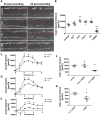
Fig 2. Regulation of intestinal epithelial wound healing by potassium channel modulation.
Wound healing of IEC-18 within six hours after mechanical injury. A: Representative images of wounds 0 and 6 hours after wounding in the presence of the indicated potassium channel modulators. Scale bar: 100μm. B: Quantitative analysis of wound healing of IEC-18 incubated with different potassium channel modulators (n = 5). While IbTx and Clt cause an increase in intestinal epithelial wound healing response, wound closure is significantly reduced after administration of 1-EBIO. C-E: Time course of wound healing after administration of IbTx, Clt and 1-EBIO (n = 5). F: Comparison of different solvent concentration applied (n = 5). While 0.25% (v/v) DMSO does not affect wound closure, 1% (v/v) DMSO significantly retards wound closure. G: Direct comparison of 1-EBIO with its solvent (n = 5). Reduction in wound healing by 1-EBIO is also significant vs. 1% DMSO.
Fig 3
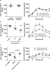
Fig 3. Differential regulation of KCNN4-dependent intestinal epithelial wound healing in response to preincubation with IFN-γ.
Wound healing of IEC-18 within six hours after mechanical injury. A: Comparison of wound healing of IEC-18 under control conditions without and with 24 h-pretreatment with IFN-γ (n = 5). Both the mean wound closure over six hours (left panel) and the time course profile of wound healing (right panel) are similar. B: Wound healing of IEC-18 after IFN-γ pretreatment and incubation with Clt and 1-EBIO (n = 5). KCNN4-inhibition by Clt results in significantly reduced rates of wound closure (left panel). Compared to Clt treatment without IFN-γ preincubation wound closure rates upon addition of Clt after 24h IFN-γ are significantly reduced in the second, third and fourth hour (right panel). C: Comparison of 1-EBIO-dependent wound closure to 1% DMSO (left panel, n = 5) and time course of 1-EBIO-dependent restitution with and without IFN-γ preincubation (right panel, n = 5). After proinflammatory treatment, 1-EBIO-dependent wound closure is higher than under baseline conditions.
Fig 4
Fig 4. Potassium channel-dependent wound healing of IEC-18 with and without 5 nm EGF under baseline or IFN-γ pretreated conditions.
Wound healing of IEC-18 within six hours after mechanical injury. A: Influence of EGF on intestinal epithelial wound healing (n = 5). EGF increases wound healing both under baseline and inflammatory conditions (left panel). Right panel: Time course of restitution upon EGF treatment compared to control showing marked differences in the second, third and fourth hour after wounding. B: Impact of Clt with and without additional EGF after or without IFN-γ pretreatment (n = 5). EGF does not further increase Clt-dependent wound healing under baseline conditions, but fully reverts the Clt-dependent reduction in wound closure after proinflammatory treatment. C: Impact of 1-EBIO with and without additional EGF after or without IFN-γ pretreatment (n = 3–5). EGF promotes 1-EBIO-mediated wound healing both under baseline and inflammatory conditions.
Fig 5
Fig 5. Boyden chamber results.
A: Representative images of stained cells attached to the lower surface of polycarbonate membranes after migration of IEC-18 towards medium containing Clt or not and after or without IFN-γ pretreatment. B: Quantitative analysis of the number of cells migrated through a polycarbonate membrane during six hours under the indicated conditions (n = 14–16). Clt-dependent migration is significantly higher under baseline than under inflammatory conditions.
Fig 6
Fig 6. Clt-dependent alterations in Akt but not ERK phosphorylation correlate with wound healing of IEC-18.
A: Representative blots showing Akt phosphorylation after 30 and 120 minutes. B: Representative blots showing ERK phosphorylation after 30 and 120 minutes. n.w.–not wounded. C-E: Quantitative analysis of the phosphorylation of Akt (left panels) and ERK (right panels) as determined by pAkt/Akt and pERK/ERK, respectively, after 30 (C), 120 (D) and 480 minutes (E). Clt enhances Akt but not ERK phosphorylation after 30 and 120 minutes. n = 3–7. F: Phosphorylation of Akt (upper panel) and ERK (lower panel) in Clt-involving conditions 120 minutes after wounding (n = 3–5). While ERK phosphorylation is not significantly altered, Akt phosphorylation is reduced by IFN-γ pretreatment and reraised by additional EGF, thus correlating Clt-dependent wound closure in scratch assays. G: Correlation of pAkt/Akt with mean wound closure in the second to fourth hour of the scratch assays in controls and Clt-involving constellations. Relative phosphorylation and wound closure rates (S1 Table) were plotted against each other and a regression line was computed. Pearson’s coefficient is 0.942 (p = 0.005).
Fig 7
Fig 7. Scratch-wounding of HT-29 cells confirms a differential regulation of KCNN4-dependent restitution with crucial contribution of PI3K but not ERK signaling.
Confluent monolayers of HT-29 cells were wounded as explained in the Methods section and imaged right after wounding and after 6h. A: Representative images of the wounds at the indicated time points upon treatment with the indicated compounds without (left panels) or after (right panels) 24h pretreatment with IFN-γ. Wound size is denoted in red. Scale bar: 100 μm. B: Quantitative analysis of wound healing under baseline conditions (left panels) or with proinflammatory treatment (right panels) and without (upper panels) or with EGF treatment (lower panels), respectively (n = 11–24). Individual wound closure is displayed as % difference vs. the mean of respective controls. Clt increases wound healing vs. controls in baseline but not inflammatory conditions. Clt in combination with EGF does not further increase EGF-dependent wound healing. C: Impact of PI3K (left panel) and ERK inhibition (right panel) on EGF-dependent intestinal epithelial restitution (n = 4–24). Both compounds revert the increase observed with EGF. D: Impact of PI3K (left panel) and ERK inhibition (right panel) on Clt-dependent intestinal epithelial wound healing (n = 4–24). The Clt-mediated increase in wound closure is abrogated by addition of the PI3K inhibitor but not the ERK inhibitor.
Fig 8
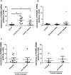
Fig 8. KCNN4 mRNA expression in IBD.
A: Relative levels of KCNN4 mRNA in IEC from controls (n = 10), UC (n = 8) and CD (n = 14). In both UC and CD the expression is increased vs. control. B: KCNQ1 mRNA levels are equal in UC (n = 5), CD (n = 11) and controls (n = 10) C: Comparison of KCNN4 expression in inflamed vs. uninflamed tissue of patients with CD. Left panel: Relative KCNN4 mRNA levels in all available samples (n = 14 inflamed, n = 7 not inflamed). Right panel: Comparison of KCNN4 levels in inflamed and uninflamed samples originating from the same patients (n = 5).
Fig 9
Fig 9. Hypothesized impact of KCNN4 inhibition on wound-induced migration and differential regulation by IFN-γ.
A: Blockade of KCNN4 by Clt (red X) accelerates wound healing at least in part by transactivation of the PI3K pathway (red arrows). Additional EGF (blue) meets activated downstream signaling and exerts no additional effect (blue arrows). B: IFN-γ preincubation uncouples the postulated transactivation of PI3K signaling by KCNN4 inhibition (orange line) leading to reduced migration upon KCNN4 blockade by Clt (red X). In this constellation EGF (blue) may activate downstream targets and enhance migration (blue arrows).

Similar articles

Cited by

References

    1. Baumgart DC, Dignass AU. Intestinal barrier function. Curr Opin Clin Nutr Metab Care. 2002;5: 685–694. - PubMed
    1. Dignass AU. Mechanisms and modulation of intestinal epithelial repair. Inflamm Bowel Dis. 2001;7: 68–77. - PubMed
    1. Polk DB. Epidermal growth factor receptor-stimulated intestinal epithelial cell migration requires phospholipase C activity. Gastroenterology. 1998;114: 493–502. - PubMed
    1. Wilson AJ, Gibson PR. Role of epidermal growth factor receptor in basal and stimulated colonic epithelial cell migration in vitro. Exp Cell Res. 1999;250: 187–196. 10.1006/excr.1999.4496 - DOI - PubMed
    1. Riegler M, Sedivy R, Sogukoglu T, Cosentini E, Bischof G, Teleky B, et al. Epidermal growth factor promotes rapid response to epithelial injury in rabbit duodenum in vitro. Gastroenterology. 1996;111: 28–36. - PubMed

Publication types

MeSH terms

Substances

LinkOut - more resources