. 2016 Jan 29;11(1):e0147389.
doi: 10.1371/journal.pone.0147389. eCollection 2016.

Therapeutic Use of 3β-[N-(N',N'-Dimethylaminoethane) Carbamoyl] Cholesterol-Modified PLGA Nanospheres as Gene Delivery Vehicles for Spinal Cord Injury

Affiliations

Therapeutic Use of 3β-[N-(N',N'-Dimethylaminoethane) Carbamoyl] Cholesterol-Modified PLGA Nanospheres as Gene Delivery Vehicles for Spinal Cord Injury

So-Jung Gwak et al. PLoS One. .

Abstract

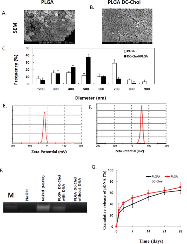
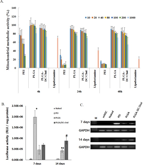
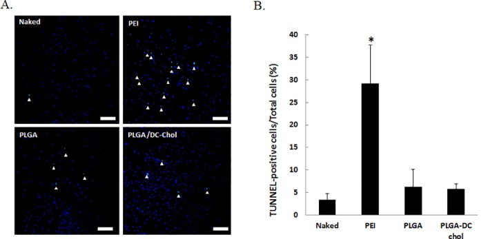
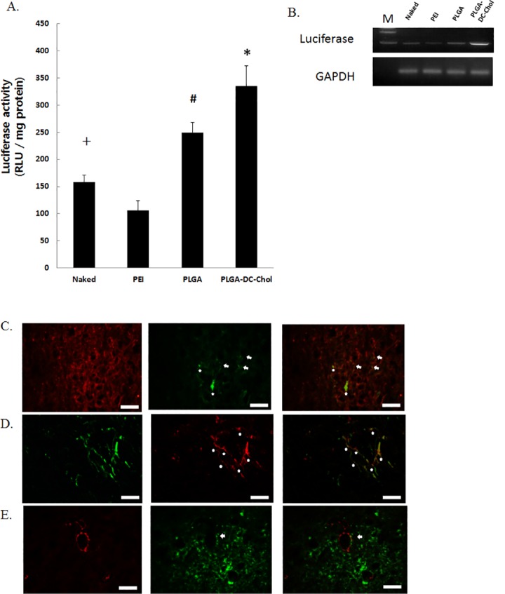
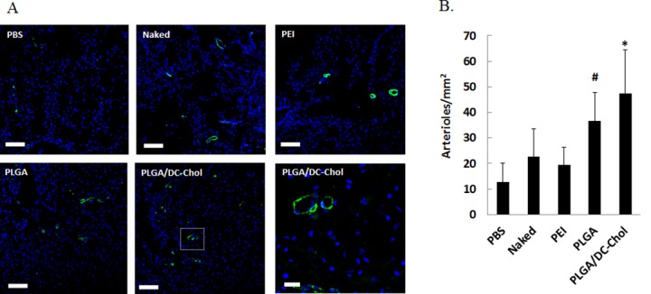
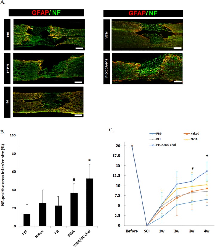
Gene delivery holds therapeutic promise for the treatment of neurological diseases and spinal cord injury. Although several studies have investigated the use of non-viral vectors, such as polyethylenimine (PEI), their clinical value is limited by their cytotoxicity. Recently, biodegradable poly (lactide-co-glycolide) (PLGA) nanospheres have been explored as non-viral vectors. Here, we show that modification of PLGA nanospheres with 3β-[N-(N',N'-dimethylaminoethane) carbamoyl] cholesterol (DC-Chol) enhances gene transfection efficiency. PLGA/DC-Chol nanospheres encapsulating DNA were prepared using a double emulsion-solvent evaporation method. PLGA/DC-Chol nanospheres were less cytotoxic than PEI both in vitro and in vivo. DC-Chol modification improved the uptake of nanospheres, thereby increasing their transfection efficiency in mouse neural stem cells in vitro and rat spinal cord in vivo. Also, transgene expression induced by PLGA nanospheres was higher and longer-lasting than that induced by PEI. In a rat model of spinal cord injury, PLGA/DC-Chol nanospheres loaded with vascular endothelial growth factor gene increased angiogenesis at the injury site, improved tissue regeneration, and resulted in better recovery of locomotor function. These results suggest that DC-Chol-modified PLGA nanospheres could serve as therapeutic gene delivery vehicles for spinal cord injury.


Conflict of interest statement

Competing Interests: The authors have declared that no competing interests exist.

Figures

Fig 1
Fig 1. Characterization of nanospheres.
SEM images of (A) PLGA and (B) PLGA/DC-Chol nanospheres. (C) Size distribution of nanospheres. Zeta potential (mV) of (D) PLGA and (E) PLGA/DC-Chol nanospheres. (F) Agarose gel retardation analysis. Agarose gel electrophoresis of pDNA released from PLGA and PLGA/DC-Chol nanospheres. (G) Cumulative release of pDNA from PLGA and PLGA/DC-Chol nanospheres.
Fig 2
Fig 2. (A) Cytotoxicity of pDNA-loaded nanospheres and PEI/pDNA complexes. mNSCs were cultured for 4, 24, or 48 h with the indicated polymer concentrations, and mitochondrial metabolic activity in mNSCs was measured using the MTT assay. p < 0.05 for nanospheres (PLGA/DC-Chol or PLGA) vs. PEI at all concentrations and time points (n = 5) except for 10 and 20 μg at 4 h.
Transfection efficiency in cultured mNSCs (B, C). (B) Luciferase gene expression in mNSCs transfected with pSV-Luc-loaded PLGA nanospheres, PLGA/DC-Chol nanospheres, or PEI /pSV-Luc complex. *p < 0.05.PEI/pLuci compare with naked, PLGA and PLGA/DC-Chol at 7 days after transfection. # p < 0.05 PLGA/DC-Chol nanospheres compared with PLGA, PEI and naked at 14 days after transfection. **p < 0.05 PLGA nanospheres compared with PEI and naked at 14 days after transfection. (C) Luciferase mRNA expression 7 and 14 days after gene transfection.
Fig 3
Fig 3. Apoptosis in the rat spinal cord.
(A) Apoptotic activity at the injection site 24 h after injection. Nuclei were stained with DAPI (blue). Apoptosis-positive nuclei were stained with FITC (green) using a TUNEL staining method. Scale bars indicate 100 μm. (B) TUNEL-positive cell density at the injection site for naked pDNA, PEI/pDNA, PLGA/pDNA, and PLGA/DC-Chol/pDNA groups. *p < 0.05 compared with naked, PLGA nanospheres and PLGA/DC-Chol nanospheres
Fig 4
Fig 4. Gene expression in the rat spinal cord.
(A, B) Luciferase activity in the rat spinal cord 14 days after injection. *p < 0.05 compared with naked pLuci and PLGA/pLuci. # p < 0.05 compared with naked and PEI/pDNA, + p < 0.05 compared with PEI/pDNA. Duble immunofluorescent staining for (C) beta III tublin (red), luciferease (green) and betaIII tublin/luciferase merged cells (yellow) (D) GFAP (green), luciferase (red) and GFAP/luciferase merged cells (yellow), (E) Smooth muscle α-actin (red), luciferase (green) and SM α-actin/luciferase merged cells(yellow) in spinal cord. The arrows indicate cells of luciferase expression and beta III tublin positive cells, GFAP positive cells or sm-α actin positive cells. Scale bars indicate 50 μm.
Fig 5
Fig 5. Angiogenesis after injection of VEGF-loaded PLGA/DC-Chol nanospheres.
(A) Immunofluorescent staining for SM-α actin in injured spinal cords 4 weeks after injection. Scale bars indicate 100 μm at 100× and 20 μm at 400× (B) Quantification of arteriole density in injured spinal cords. *p < 0.05 vs. PBS, naked VEGF, or PEI/VEGF. #p < 0.05 vs. PBS.
Fig 6
Fig 6. Axonal growth at lesion site and functional recovery 4 weeks after injury and injection.
(A) Double immunostaining with GFAP (red) and NF (green). Scare bar indicates 100 μm. (B) Quantification of NF optical density of regenerated area. *p < 0.05 vs. PBS, naked VEGF, and PEI/VEGF. #p < 0.05 vs. PBS. (C) Effect of VEGF-loaded PLGA/DC-Chol nanospheres on recovery of locomotor function. BBB score was significantly greater in the PLGA/DC-Chol nanosphere group (n = 5) compared to PLGA/VEGF (n = 5), PEI/VEGF (n = 5), naked VEGF (n = 5), and PBS (n = 5) 3 and 4 weeks after injury. *p < 0.05 for PLGA/DC-Chol vs. PBS, naked VEGF, and PEI/VEGF groups.

References

    1. Federici T, Boulis NM. Gene-based treatment of motor neuron diseases. Muscle Nerve. 2006;33: 302–323. - PubMed
    1. Blits B, Bunge MB. Direct gene therapy for repair of the spinal cord. J Neurotrauma. 2006;23: 508–520. - PubMed
    1. Mandel RJ, Spratt SK, Snyder RO, Leff SE. Midbrain injection of recombinant adeno-associated virus encoding rat glial cell line-derived neurotrophic factor protects nigral neurons in a progressive 6-hydroxydopamine-induced degeneration model of Parkinson's disease in rats. Proc Natl Acad Sci U S A. 1997;94: 14083–14088. - PMC - PubMed
    1. Frampton AR Jr, Goins WF, Nakano K, Burton EA, Glorioso JC. HSV trafficking and development of gene therapy vectors with applications in the nervous system. Gene Ther. 2005;12: 891–901. - PubMed
    1. Griesenbach U, Meng C, Farley R, Wasowicz MY, Munkonge FM, Chan M, et al. The use of carboxymethylcellulose gel to increase non-viral gene transfer in mouse airways. Biomaterials. 2010;31: 2665–2672. 10.1016/j.biomaterials.2009.12.005 - DOI - PMC - PubMed

Publication types

MeSH terms

Substances