. 2016 Jan 29;11(1):e0148018.
doi: 10.1371/journal.pone.0148018. eCollection 2016.

Histatin-1 Expression in Human Lacrimal Epithelium

Affiliations

Histatin-1 Expression in Human Lacrimal Epithelium

Dhara Shah et al. PLoS One. .

Abstract

Background: Study of human lacrimal cell biology is limited by poor access to tissue samples, heterogeneous cell composition of tissue and a lack of established lacrimal epithelial markers. In order to further our understanding of lacrimal cell biology, we sought to find a better marker for human lacrimal epithelial cells, compared to what has been reported in the literature.

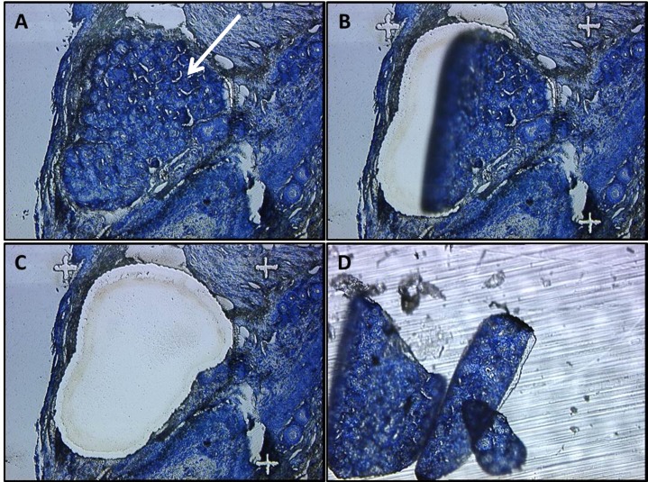
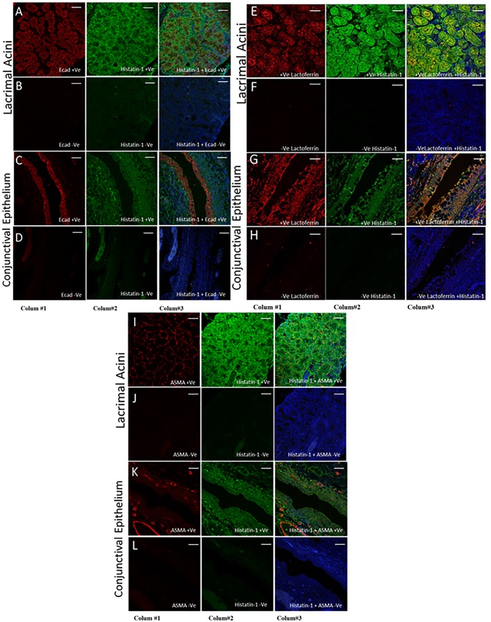
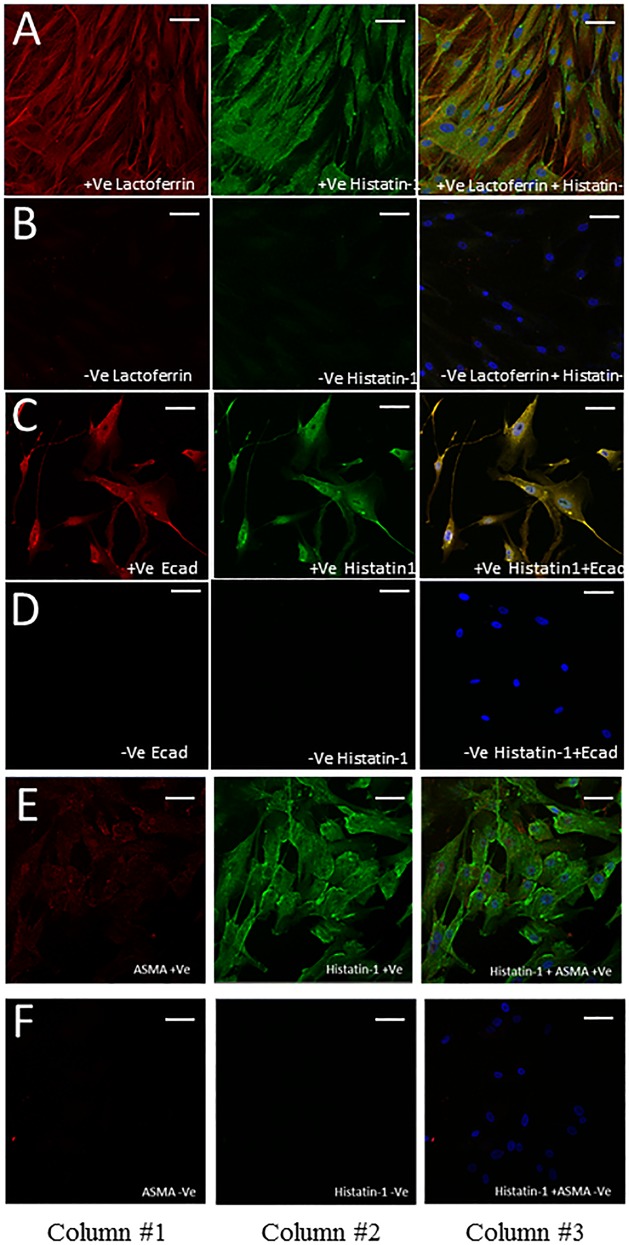
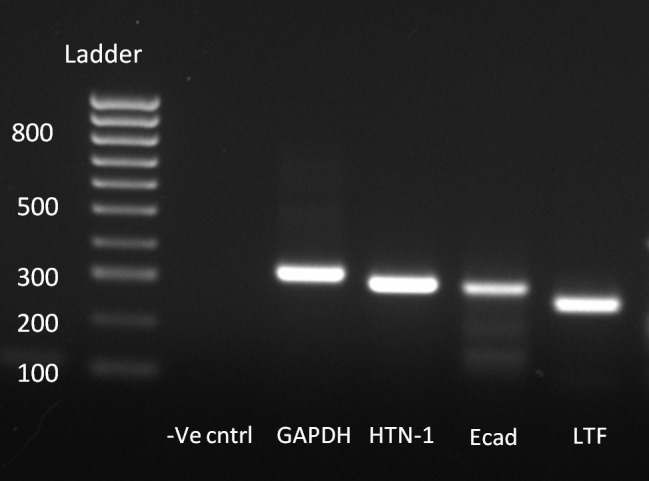
Methods: We utilized human Muller's muscle conjunctival resection (MMCR) specimens containing accessory lacrimal gland (ALG) and cadaveric main lacrimal gland (MLG) as sources of lacrimal tissue. Candidate markers were sought using human ALG tissue from MMCR specimens, isolated by laser capture microdissection (LCM). Affymetrix® analysis was performed on total RNA isolated from FFPE samples to profile transcription in ALG. MMCR tissue sections were assessed by immunofluorescence using antibodies for histatin-1, lactoferrin, E-cadherin (E-cad) and alpha-smooth muscle actin (ASMA). Reverse transcriptase polymerase chain reaction (RT-PCR) analysis was performed to analyze the expression of histatin-1, E-cad and lactoferrin from cadaveric MLG.

Results: Histatin-1 is expressed in ALG and MLG, localizes to lacrimal epithelium, and to a greater degree than do other putative lacrimal epithelial markers.

Conclusions: Histatin-1 is a good marker for human lacrimal epithelium in ALG and MLG and can be used to identify lacrimal cells in future studies.


Conflict of interest statement

Competing Interests: The authors have declared that no competing interests exist.

Figures

Fig 1
Fig 1. LASER Capture Microdissection of Accessory Lacrimal Gland.
LCM of ALG from FFPE Human MMCR specimens. (A) Intact ALG in an MMCR specimen (white arrow). (B, C) LASER dissection of ALG from MMCR D. Isolated ALG tissue.
Fig 2
Fig 2. Immunofluorescence of ALG Specific Markers in MMCR.
Immunofluorescence staining of ALG specific markers in MMCR specimens showing the difference in localization of markers (Ecad, Histatin-1, lactoferrin and ASMA) between lacrimal acini and conjunctival epithelium. Column 1 depicts a single stain for E-cad (Rows A-D), Lactoferrin (Rows E-H), and ASMA (Rows I-L). Column 2 shows stain for histatin-1. Column 3 shows an overlapped image of Column 1 and 2 and blue staining for nuclei (DAPI). Rows B, D, F, H, J and L are negative control images. Rows A-D show strong localization of E-cad and histatin-1 in lacrimal acini, with strong overlap and lesser localization of histatin-1 to conjunctival epithelium than lacrimal acini. In contrast, E-cad localizes to conjunctival epithelium better than it does lacrimal acini. Rows E-H show localization of lactoferrin and histatin-1 to lacrimal acini, with strong overlap and lesser localization of histatin-1 to conjunctival epithelium than to lacrimal acini. In contrast, lactoferrin localizes to conjunctival epithelium better than it does lacrimal acini. Rows I-J show good localization of ASMA to myoepithelial cells surrounding lacrimal acini, and good localization of histatin-1 to lacrimal acini without significant overlap. Rows K and L demonstrate no localization of ASMA to conjunctival epithelium by ASMA, and low levels of localization of histatin-1 to conjunctival epithelium. Scale bar, 50 μm.
Fig 3
Fig 3. Immunofluorescence staining of cultured MLG cells.
Immunofluorescence staining in cultured MLG cells using antibodies to lactoferrin (Row A), E-cad (Row C), ASMA (Row E) and histatin-1 (Rows A, C, E). Rows B, D and F are respective negative control images. Column 1 depicts a single stain for antibodies to lactoferrin (Rows A) E-cad (Rows C, D), ASMA (Rows E,F). Column 2 shows stain for histatin-1. Column 3 shows an overlapped image of Column 1 and 2 and blue staining for nuclei (DAPI). Row A shows strong localization of lactoferrin and histatin-1 to cultured MLG epithelium. Row C shows strong localization of E-cad and histatin-1 to cultured MLG epithelium. Row E shows strong localization of histain-1 to cultured MLG epithelium but ASMA does not localize well to cultured MLG epithelium. Scale bar, 50 μm.
Fig 4
Fig 4. RT-PCR analysis of human main lacrimal gland tissue.
RT-PCR of main lacrimal gland tissue shows expression of histatin-1, purported lacrimal markers E-cad and lactoferrin.

References

    1. Brewitt H, Sistani F. Dry eye disease: the scale of the problem. Surv Ophthalmol. 2001;45 Suppl 2:S199–202. Review - PubMed
    1. Tiwari S, Ali MJ, Vemuganti GK. Human lacrimal gland regeneration: Perspectives and review of literature. Saudi J Ophthalmol. 2014;1:12–8. - PMC - PubMed
    1. Liu A, Cheng L, Du J, Peng Y, Allan RW, Wei L et al. Diagnostic utility of novel stem cell markers SALL4, OCT4, NANOG, SOX2, UTF1, and TCL1 in primary mediastinal germ cell tumors. Am J Surg Pathol. 2010;34(5):697–706. 10.1097/PAS.0b013e3181db84aa - DOI - PubMed
    1. Tiwari S, Ali MJ, Balla MM, Naik MN, Honavar SG, Reddy VA, et al. Establishing human lacrimal gland cultures with secretory function. PLoS One. 2012;7(1):e29458 10.1371/journal.pone.0029458 Epub 2012 Jan 13. - DOI - PMC - PubMed
    1. Spaniol K, Metzger M, Roth M, Greve B, Mertsch S, Geerling G, et al. Engineering of a secretory active three-dimensional lacrimal gland construct on the basis of decellularized lacrimal gland tissue. Tissue Eng Part A. 2015;21(19–20):2605–17. 10.1089/ten.TEA.2014.0694 - DOI - PubMed

Publication types

MeSH terms