Ossifying fibromas of the paranasal sinuses: diagnosis and management
- PMID: 26824919
- PMCID: PMC4720932
- DOI: 10.14639/0392-100X-533
Ossifying fibromas of the paranasal sinuses: diagnosis and management
Abstract
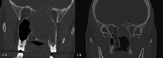
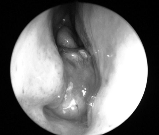
Fibro-osseous benign lesions rarely affect the sinonasal tract and are divided into 3 different entities, namely osteoma, fibrous dysplasia and ossifying fibroma. They share several clinical, radiological and histological similarities, but have different behaviours. Ossifying fibroma, and in particular the "juvenile" histological subtype, may have a locally aggressive evolution and a high risk for recurrence if removal is incomplete. The purpose of the present study is to compare the clinical behaviour of ossifying fibroma with the other benign fibro-osseous lesions; highlight different behaviour between the histological subtypes; compare the advantages, limitations and outcomes of an endoscopic endonasal approach with reports in the literature. We retrospectively reviewed 11 patients treated for sinonasal ossifying fibroma at a tertiary care centre. All patients underwent CT scan, and MRI was performed in cases of cranial base involvement or recurrence. Pre-operative biopsy was performed in cases where it was possible to use an endoscopic approach. One patient underwent pre-operative embolisation with ipsilateral visual loss after the procedure. Depending on its location, removal of the tumour was performed using an endoscopic (n = 7), or an external (n = 3) or combined (n = 1) approach. Histopathologically, 5 patients presented the conventional type, 5 the juvenile psammomatoid variant, which was associated in 1 case with an aneurismal bone cyst, and 1 case presented the trabecular juvenile variant. Three patients affected by the juvenile psammomatoid histological variant presented invasion of the skull base and underwent a subtotal removal that subsequently required, due to the regrowth of the remnant, a transbasal approach. Clinical, radiological and histological findings should all be considered to establish differential diagnosis among fibrous osseous lesions. More studies are necessary to conclude if the localisation and extension of the disease at the time of diagnosis is more important than the histological variant. An endoscopic approach is the first choice in most of cases even if an external open approach may be necessary in selected patients.
Le lesioni fibro-ossee benigne raramente colpiscono i seni paranasali e sono suddivise in 3 entità: osteoma, displasia fibrosa e fibroma ossificante. Questi presentano simili caratteristiche cliniche, radiologiche e istologiche ma hanno un comportamento diverso. Il fibroma ossificante, in particolare la variante istologica giovanile, può presentare un comportamento aggressivo con un alto rischio di recidiva se rimosso in modo incompleto. Lo scopo dello studio è quello di paragonare il comportamento clinico del fibroma ossificante con quello delle altre lesioni fibro-ossee; di evidenziare un eventuale comportamento differente tra i vari sottotipi istologici; di descrivere i vantaggi, i limiti e i risultati della chirurgia endoscopica endonasale rispetto ai dati presenti in letteratura. Abbiamo analizzato retrospettivamente 11 pazienti affetti da fibroma ossificante naso-sinusale e trattati in un centro ospedaliero di terzo livello. Tutti i pazienti sono stati sottoposti a TC. La RM è stata eseguita in caso di coinvolgimento del basicranio o in caso di recidiva. Una biopsia pre-operatoria è stata effettuata nei casi in cui la massa era raggiungibile per via endoscopica. Un paziente è stato sottoposto a embolizzazione pre-operatoria ed ha riportato una cecità monolaterale al termine della procedure. In base alla localizzazione, l'exeresi del tumore è stata eseguita con un approccio endoscopico (7 pazienti), esterno (3), combinato (1). Istologicamente 5 pazienti hanno riportato un sottotipo convenzionale, 5 la variante giovanile psammomatoide associata in un caso a cisti aneurismatica ossea, e un paziente la variante giovanile trabecolare. Tre pazienti affetti dalla variante istologica giovanile psammomatoide hanno presentato un'invasione del basicranio e sono stati sottoposti ad exeresi subtotale per via endoscopica che ha richiesto in seguito, a causa di un aumento di volume del residuo, un secondo intervento per via transbasale. I reperti clinici, radiologici e istologici dovrebbero essere considerati insieme per una accurata diagnosi differenziale tra le lesioni fibro-ossee. Ulteriori studi sono necessari per concludere se la localizzazione e l'estensione del fibroma ossificante al momento della diagnosi sono più importanti della variante istologica. L'approccio endoscopico è la prima opzione nella maggior parte dei casi anche se in alcuni selezionati pazienti l'approccio esterno risulta ancora necessario.
Keywords: Endoscopic surgery; Fibrous dysplasia; Ossifying fibroma; Osteoma; Skull base.
Figures
Similar articles
-
Chromosome 12 long arm rearrangement covering MDM2 and RASAL1 is associated with aggressive craniofacial juvenile ossifying fibroma and extracranial psammomatoid fibro-osseous lesions.Mod Pathol. 2015 Jan;28(1):48-56. doi: 10.1038/modpathol.2014.80. Epub 2014 Jun 13. Mod Pathol. 2015. PMID: 24925056
-
[Surgical management of sinonasal and adjacent skull base benign fibro-osseous lesions].Lin Chuang Er Bi Yan Hou Tou Jing Wai Ke Za Zhi. 2011 Mar;25(5):226-31. Lin Chuang Er Bi Yan Hou Tou Jing Wai Ke Za Zhi. 2011. PMID: 21604470 Chinese.
-
Aggressive psammomatoid ossifying fibromas of the sinonasal region: a clinicopathologic study of a distinct group of fibro-osseous lesions.Cancer. 1995 Oct 1;76(7):1155-65. doi: 10.1002/1097-0142(19951001)76:7<1155::aid-cncr2820760710>3.0.co;2-p. Cancer. 1995. PMID: 8630892
-
Juvenile psammomatoid ossifying fibroma: a review.Oral Oncol. 2011 Dec;47(12):1110-6. doi: 10.1016/j.oraloncology.2011.06.513. Epub 2011 Aug 12. Oral Oncol. 2011. PMID: 21840246 Review.
-
Common fibro-osseous lesions of the paranasal sinuses.Otolaryngol Clin North Am. 2006 Jun;39(3):585-600, x. doi: 10.1016/j.otc.2006.01.013. Otolaryngol Clin North Am. 2006. PMID: 16757233 Review.
Cited by
-
Giant Post-Traumatic Frontoethmoid Osteoma: Diagnostic, Therapeutic and Reconstructive Approach.Turk Arch Otorhinolaryngol. 2020 Mar;58(1):61-64. doi: 10.5152/tao.2020.4858. Epub 2020 Mar 1. Turk Arch Otorhinolaryngol. 2020. PMID: 32313898 Free PMC article.
-
Juvenile psammomatoid ossifying fibroma: A radiolucent lesion to suspect preoperatively.Radiol Case Rep. 2019 Jun 8;14(8):1014-1020. doi: 10.1016/j.radcr.2019.05.017. eCollection 2019 Aug. Radiol Case Rep. 2019. PMID: 31198486 Free PMC article.
-
Pediatric Benign Fibro-Osseous Lesions of the Nose and Paranasal Sinuses: A Tertiary Hospital Experience.Int J Otolaryngol. 2022 Aug 25;2022:1608015. doi: 10.1155/2022/1608015. eCollection 2022. Int J Otolaryngol. 2022. PMID: 36060197 Free PMC article.
-
Unilateral visual loss resulting from orbital encroachment of an ethmoidal juvenile trabecular ossifying fibroma.Ann R Coll Surg Engl. 2019 Apr;101(4):e111-e114. doi: 10.1308/rcsann.2019.0030. Epub 2019 Mar 11. Ann R Coll Surg Engl. 2019. PMID: 30854866 Free PMC article.
-
A practical overview of CT and MRI features of developmental, inflammatory, and neoplastic lesions of the sphenoid body and clivus.Neuroradiology. 2022 Aug;64(8):1483-1509. doi: 10.1007/s00234-022-02986-x. Epub 2022 Jun 3. Neuroradiology. 2022. PMID: 35657394 Free PMC article. Review.
References
-
- Speight PM, Carlos R. Maxillofacial fibro-osseous lesions. Curr Diagn Pathol. 2006;12:1–10.
-
- Commins DJ, Tolley NS, Milford CA. Fibrous dysplasia and ossifying fibroma of the paranasal sinuses. J Laryngol Otol. 1998;112:964–968. - PubMed
-
- Eller R, Sillers M. Common fibro-osseous lesions of the paranasal sinuses. Otolaryngol Clin North Am. 2006;39:585–600. - PubMed
-
- Lund VJ, Stammberger H, Nicolai P, et al. European position paper on endoscopic management of tumours of the nose, paranasal sinuses and skull base. Rhinol Suppl. 2010;22:1–143. - PubMed
-
- Ledderose GJ, Stelter K, Becker S, et al. Paranasal ossifying fibroma: endoscopic resection or wait and scan? Eur Arch Otorhinolaryngol. 2011;268:999–1004. - PubMed
MeSH terms
LinkOut - more resources
Full Text Sources
Medical