. 2016 Mar 25;60(4):2028-38.
doi: 10.1128/AAC.02221-15. Print 2016 Apr.

Nitazoxanide Inhibits Pilus Biogenesis by Interfering with Folding of the Usher Protein in the Outer Membrane

Affiliations

Nitazoxanide Inhibits Pilus Biogenesis by Interfering with Folding of the Usher Protein in the Outer Membrane

Peter Chahales et al. Antimicrob Agents Chemother. .

Abstract

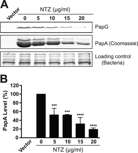
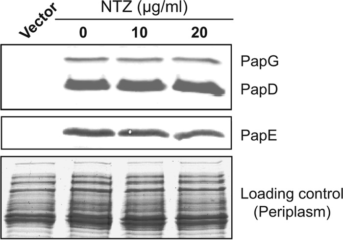
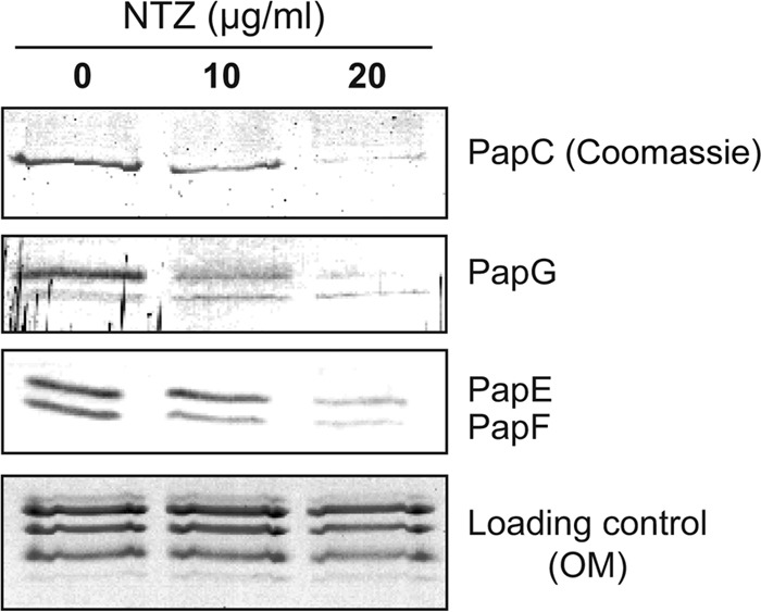
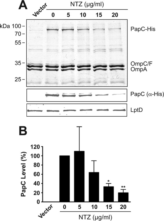
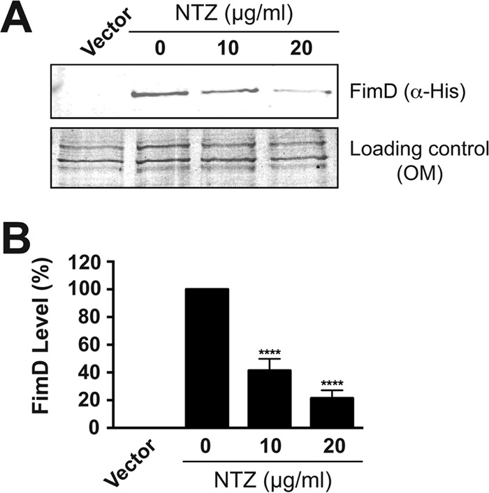
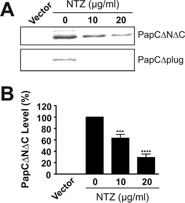
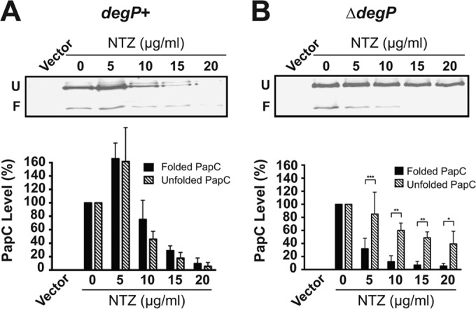
Many bacterial pathogens assemble surface fibers termed pili or fimbriae that facilitate attachment to host cells and colonization of host tissues. The chaperone/usher (CU) pathway is a conserved secretion system that is responsible for the assembly of virulence-associated pili by many different Gram-negative bacteria. Pilus biogenesis by the CU pathway requires a dedicated periplasmic chaperone and an integral outer membrane (OM) assembly and secretion platform termed the usher. Nitazoxanide (NTZ), an antiparasitic drug, was previously shown to inhibit the function of aggregative adherence fimbriae and type 1 pili assembled by the CU pathway in enteroaggregativeEscherichia coli, an important causative agent of diarrhea. We show here that NTZ also inhibits the function of type 1 and P pili from uropathogenicE. coli(UPEC). UPEC is the primary causative agent of urinary tract infections, and type 1 and P pili mediate colonization of the bladder and kidneys, respectively. By analysis of the different stages of the CU pilus biogenesis pathway, we show that treatment of bacteria with NTZ causes a reduction in the number of usher molecules in the OM, resulting in a loss of pilus assembly on the bacterial surface. In addition, we determine that NTZ specifically prevents proper folding of the usher β-barrel domain in the OM. Our findings demonstrate that NTZ is a pilicide with a novel mechanism of action and activity against diverse CU pathways. This suggests that further development of the NTZ scaffold may lead to new antivirulence agents that target the usher to prevent pilus assembly.


Figures

FIG 1
FIG 1
Effect of NTZ on P pilus assembly on the bacterial surface. (A) Strain AAEC185/pJF29 was grown in the presence of increasing concentrations of NTZ and was induced for expression of P pili. Pili isolated from the bacterial surface were separated by SDS-PAGE and were blotted with anti-PapD-PapG antibody to visualize the PapG adhesin (top panel) or stained with Coomassie blue to visualize the major pilus subunit PapA (middle panel). The bottom panel shows the Coomassie-stained whole bacteria used for pilus isolation as a loading control. E. coli containing vector only (pACYC184) served as a negative control for pilus isolation. (B) Quantitation of PapA levels in the isolated pili. PapA levels were measured by densitometry of the middle section of panel A, and the percentages of PapA levels were calculated relative to 0 μg/ml NTZ. Bars represent means ± standard errors of the means (SEM) from three independent experiments. ***, P < 0.001; ****, P < 0.0001 for comparison with 0 μg/ml NTZ.
FIG 2
FIG 2
Effect of NTZ on chaperone-subunit interactions in the periplasm. Strain BW25113/pPAP58 was grown in the presence of increasing concentrations of NTZ and was induced for expression of the PapD chaperone and P pilus tip subunits (PapG, E, F, and K). Periplasm fractions isolated from the bacteria were separated by SDS-PAGE and were blotted with anti-PapD-PapG antibody (top panel) to visualize the chaperone and adhesin or with anti-P pilus tip antibody to visualize the PapE major tip subunit (middle panel). The bottom panel shows the periplasm fractions stained with Coomassie blue as a loading control.
FIG 3
FIG 3
Effect of NTZ on formation of usher-chaperone-subunit complexes in bacteria. Strain BW25113ΔompT was grown in the presence of increasing concentrations of NTZ and was induced for expression of the His-tagged PapC usher (pMJ3), along with the PapD chaperone and the P pilus tip subunits (pPAP58). PapC-His, together with any stably bound pilus assembly complexes, was purified from solubilized OM fractions by nickel affinity chromatography and was separated by SDS-PAGE. The purified PapC was visualized by Coomassie blue staining (top panel). Copurifying pilus tip subunits were visualized by blotting with anti-P pilus tip antibody to detect the PapG, PapE, and PapF tip subunits (middle panels). The bottom panel shows the solubilized OM fractions stained with Coomassie blue as a loading control.
FIG 4
FIG 4
Effect of NTZ on levels of the PapC usher in the OM. (A) Strain BW25113/pMJ3 was grown in the presence of increasing concentrations of NTZ and was induced for expression of the His-tagged PapC usher. OM fractions isolated from the bacteria were subjected to SDS-PAGE and Coomassie staining to observe PapC and the major OM protein constituents (top panel). Samples were also probed with anti-His antibody to visualize PapC (middle panel) or anti-LptD antibody to visualize the LPS transporter LptD (bottom panel). E. coli containing vector only (pMON6235Δcat) served as a negative control for PapC expression. (B) Quantitation of PapC levels in the OM. PapC levels were measured by densitometry of the anti-His blot in panel A, and the percentages of PapC levels were calculated relative to 0 μg/ml NTZ. Bars represent means ± SEM from three independent experiments. *, P < 0.05; **, P < 0.01 for comparison with 0 μg/ml NTZ.
FIG 5
FIG 5
Effect of NTZ on levels of the FimD usher in the OM. (A) Strain BW25113/pETS4 was grown in the presence of increasing concentrations of NTZ and was induced for expression of the His-tagged FimD usher. OM fractions isolated from the bacteria were subjected to SDS-PAGE and were blotted with anti-His antibody to visualize FimD (top panel). The bottom panel shows the OM fractions stained with Coomassie blue as a loading control. E. coli containing vector only (pMMB66) served as a negative control for FimD expression. (B) Quantitation of FimD levels in the OM. FimD levels were measured by densitometry of the anti-His blot in panel A, and the percentages of FimD levels were calculated relative to 0 μg/ml NTZ. Bars represent means ± SEM from three independent experiments. ****, P < 0.0001 for comparison with 0 μg/ml NTZ.
FIG 6
FIG 6
Analysis of domain deletion mutants of the PapC usher. (A) Strain BW25113 transformed with plasmid pDG2ΔN2ΔC640 or pNH281 was grown in the presence of increasing NTZ concentrations and was induced for expression of PapC lacking the N- and C-terminal periplasmic domains (ΔNΔC) or the plug domain (Δplug), respectively. OM fractions isolated from the bacteria were subjected to SDS-PAGE and were blotted with anti-His antibody to visualize PapCΔNΔC (upper panel) or PapCΔplug (lower panel). E. coli containing vector only (pMON6235Δcat) served as a negative control for PapC expression. (B) Quantitation of PapCΔNΔC levels in the OM. PapC levels were measured by densitometry of the upper section of panel A, and the percentages of PapC levels were calculated relative to 0 μg/ml NTZ. Bars represent means ± SEM from three independent experiments. ***, P < 0.001; ****, P < 0.0001 for comparison with 0 μg/ml NTZ.
FIG 7
FIG 7
Analysis of usher folding in the OM. Strains BW25113 (A) or BW25113ΔdegP (B) transformed with pMJ3 were grown in the presence of increasing concentrations of NTZ and were induced for expression of the His-tagged PapC usher. OM fractions isolated from the bacteria were incubated in SDS sample buffer at 25°C, subjected to SDS-PAGE, and probed with anti-His antibody to visualize PapC. Positions of the folded (F) and unfolded (U) PapC species are indicated on the left of each gel image. E. coli containing vector only (pMON6235Δcat) served as a negative control for PapC expression. Quantitation of the folded and unfolded PapC bands is presented below each gel image. The PapC levels were measured by densitometry of the anti-His blots, and the percentages of PapC levels were calculated relative to the respective folded or unfolded species present at 0 μg/ml NTZ. Bars represent means ± SEM from three independent experiments. *, P < 0.05; **, P < 0.01; ***, P < 0.001 for comparison of folded to unfolded PapC at each NTZ concentration.

Similar articles

Cited by

References

    1. Thanassi DG, Bliska JB, Christie PJ. 2012. Surface organelles assembled by secretion systems of Gram-negative bacteria: diversity in structure and function. FEMS Microbiol Rev 36:1046–1082. doi:10.1111/j.1574-6976.2012.00342.x. - DOI - PMC - PubMed
    1. Kline KA, Falker S, Dahlberg S, Normark S, Henriques-Normark B. 2009. Bacterial adhesins in host-microbe interactions. Cell Host Microbe 5:580–592. doi:10.1016/j.chom.2009.05.011. - DOI - PubMed
    1. Sauer FG, Mulvey MA, Schilling JD, Martinez JJ, Hultgren SJ. 2000. Bacterial pili: molecular mechanisms of pathogenesis. Curr Opin Microbiol 3:65–72. doi:10.1016/S1369-5274(99)00053-3. - DOI - PubMed
    1. Cusumano CK, Hultgren SJ. 2009. Bacterial adhesion—a source of alternate antibiotic targets. IDrugs 12:699–705. - PubMed
    1. Steadman D, Lo A, Waksman G, Remaut H. 2014. Bacterial surface appendages as targets for novel antibacterial therapeutics. Future Microbiol 9:887–900. doi:10.2217/fmb.14.46. - DOI - PubMed

Publication types

MeSH terms

LinkOut - more resources