Internal carotid artery stenosis with persistent primitive hypoglossal artery treated with carotid artery stenting: A case report and literature review
- PMID: 26825135
- PMCID: PMC4978312
- DOI: 10.1177/1971400915626427
Internal carotid artery stenosis with persistent primitive hypoglossal artery treated with carotid artery stenting: A case report and literature review
Abstract
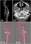
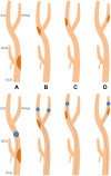
Persistent primitive hypoglossal artery (PPHA) is a persistent carotid-basilar anastomosis. It rarely remains at birth. It occasionally may be a risk for ischemia and embolic infarction to the posterior cerebral circulation, especially in patients with carotid stenosis proximal to the origin of persistent primitive arteries. We describe a case of a 60-year-old woman with asymptomatic internal carotid artery (ICA) stenosis and ipsilateral PPHA successfully treated by carotid artery stenting (CAS). A few cases of CAS for ICA stenosis with PPHA have been reported, but the strategy and methods in each case were different because of its unique anatomy and hemodynamics. It is essential to prevent distal embolisms and preserve blood flow at the territory of both the ICA and PPHA. The protection method should be selected carefully. We review the literature and discuss appropriate treatment strategies.
Keywords: Carotid artery stenosis; carotid artery stenting; cerebral embolism; embolic protection devices; persistent primitive hypoglossal artery.
© The Author(s) 2016.
Figures


Similar articles
-
Stenting for Internal Carotid Artery Stenosis Associated with Persistent Primitive Hypoglossal Artery Using Proximal Flow Blockade and Distal Protection System: A Technical Case Report and Literature Review.J Stroke Cerebrovasc Dis. 2016 Jun;25(6):e98-e102. doi: 10.1016/j.jstrokecerebrovasdis.2016.03.026. Epub 2016 Apr 19. J Stroke Cerebrovasc Dis. 2016. PMID: 27105567 Review.
-
Concomitant asymptomatic internal carotid artery and persistent primitive hypoglossal artery stenosis treated by endovascular stenting with proximal embolic protection.J Vasc Surg. 2016 Jan;63(1):237-40. doi: 10.1016/j.jvs.2014.04.066. Epub 2014 May 28. J Vasc Surg. 2016. PMID: 24877853
-
Double embolic protection during carotid artery stenting with persistent hypoglossal artery.J Neurointerv Surg. 2014 Apr 1;6(3):e23. doi: 10.1136/neurintsurg-2013-010709.rep. Epub 2013 May 9. J Neurointerv Surg. 2014. PMID: 23661691
-
A successful treatment with carotid arterial stenting for symptomatic internal carotid artery severe stenosis with ipsilateral persistent primitive hypoglossal artery: case report and review of the literature.Minim Invasive Neurosurg. 2008 Oct;51(5):298-302. doi: 10.1055/s-0028-1082299. Epub 2008 Oct 14. Minim Invasive Neurosurg. 2008. PMID: 18855296 Review.
-
Double embolic protection during carotid artery stenting with persistent hypoglossal artery.BMJ Case Rep. 2013 May 2;2013:bcr2013010709. doi: 10.1136/bcr-2013-010709. BMJ Case Rep. 2013. PMID: 23645663 Free PMC article.
Cited by
-
Stenting of the artery of Dr A.N. Kazantsev in the acute period of ischemic stroke.Radiol Case Rep. 2022 Aug 1;17(10):3699-3708. doi: 10.1016/j.radcr.2022.07.034. eCollection 2022 Oct. Radiol Case Rep. 2022. PMID: 35942267 Free PMC article.
-
Percutaneous transluminal angioplasty for persistent primitive hypoglossal artery stenosis: illustrative case.J Neurosurg Case Lessons. 2023 Oct 23;6(17):CASE23427. doi: 10.3171/CASE23427. Print 2023 Oct 23. J Neurosurg Case Lessons. 2023. PMID: 37871338 Free PMC article.
-
Stenting for elderly patients with internal carotid artery stenosis: analysis of clinical efficacy.Am J Transl Res. 2022 Oct 15;14(10):7128-7134. eCollection 2022. Am J Transl Res. 2022. PMID: 36398267 Free PMC article.
-
Bilateral persistent primitive hypoglossal arteries associated with unilateral symptomatic carotid thromboembolism.J Radiol Case Rep. 2017 Apr 30;11(4):1-9. doi: 10.3941/jrcr.v11i4.3010. eCollection 2017 Apr. J Radiol Case Rep. 2017. PMID: 28567180 Free PMC article.
-
Strategy of cerebral endovascular treatment for cervical internal carotid artery stenosis with a persistent primitive hypoglossal artery.Surg Neurol Int. 2023 Sep 1;14:308. doi: 10.25259/SNI_567_2023. eCollection 2023. Surg Neurol Int. 2023. PMID: 37810314 Free PMC article.
References
-
- De Caro R, Parenti A, Munari PF. The persistent primitive hypoglossal artery: A rare anatomic variation with frequent clinical implications. Ann Anat 1995; 177: 193–198. - PubMed
-
- Zhang L, Song G, Chen L, et al. Concomitant asymptomatic internal carotid artery and persistent primitive hypoglossal artery stenosis treated by endovascular stenting with proximal embolic protection. J Vasc Surg. Epub ahead of print 27 May 2014. DOI: 10.1016/j.jvs.2014.04.066. - PubMed
-
- Nii K, Aikawa H, Tsutsumi M, et al. Carotid artery stenting in a patient with internal carotid artery stenosis and ipsilateral persistent primitive hypoglossal artery presenting with transient ischemia of the vertebrobasilar system: Case report. Neurol Med Chir (Tokyo) 2010; 50: 921–924. - PubMed
-
- Pyun HW, Lee DH, Kwon SU, et al. Internal carotid artery stenosis with ipsilateral persistent hypoglossal artery presenting as a multiterritorial embolic infarction: A case report. Acta Radiol 2007; 48: 116–118. - PubMed
Publication types
MeSH terms
LinkOut - more resources
Full Text Sources
Other Literature Sources
Miscellaneous

