. 2016;15(2):283-94.
doi: 10.1080/15384101.2015.1100776.

Two different specific JNK activators are required to trigger apoptosis or compensatory proliferation in response to Rbf1 in Drosophila

Affiliations

Two different specific JNK activators are required to trigger apoptosis or compensatory proliferation in response to Rbf1 in Drosophila

Amandine Clavier et al. Cell Cycle. 2016.

Abstract

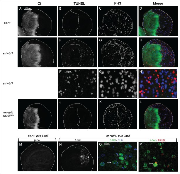
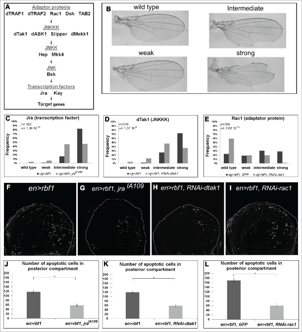
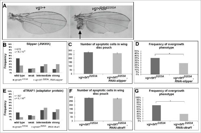
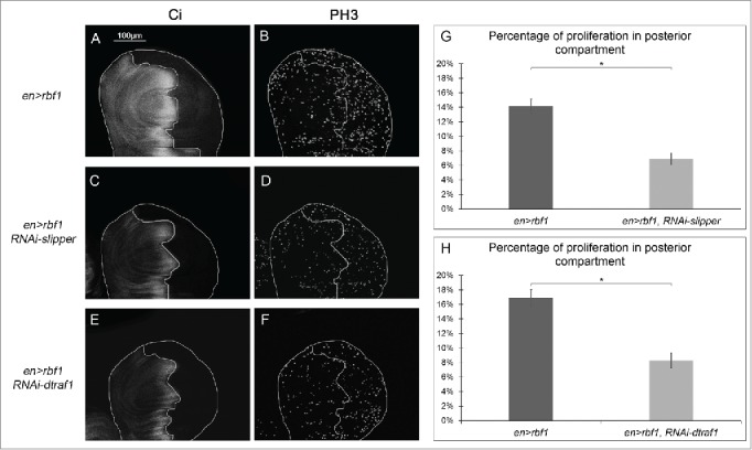
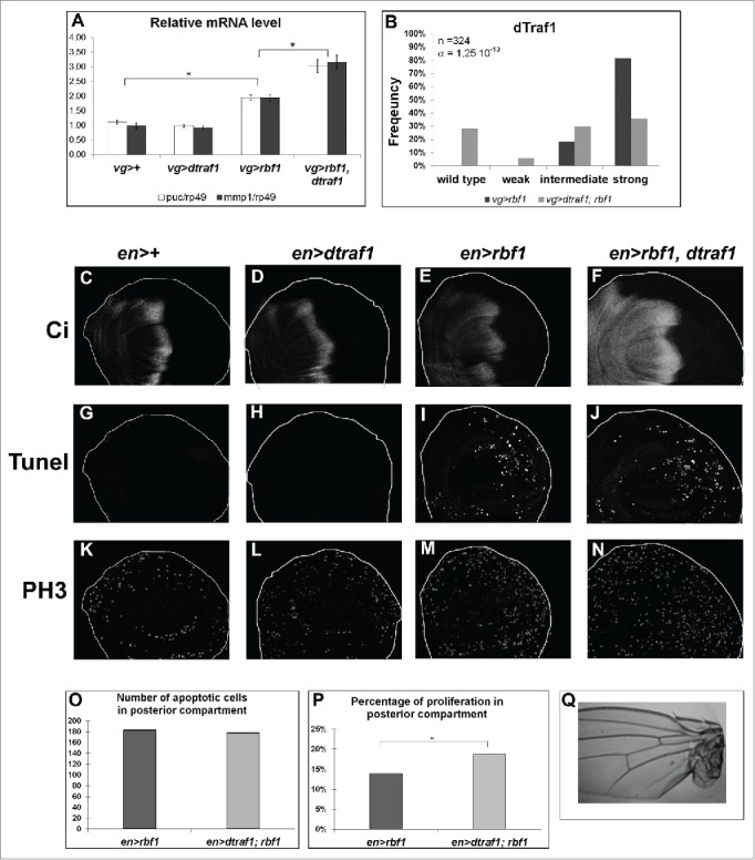
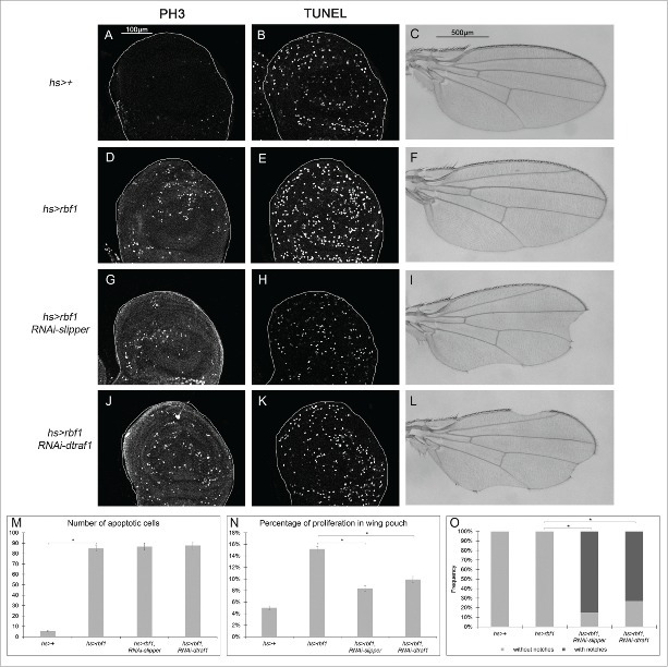
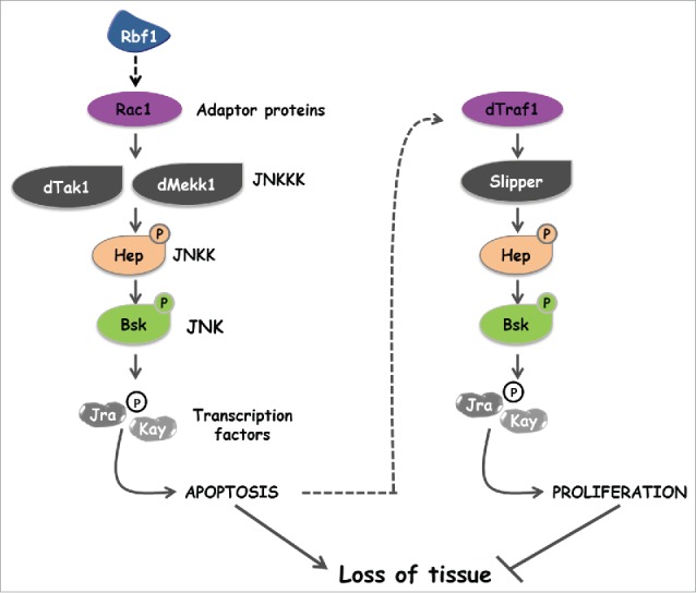
The Jun Kinase (JNK) signaling pathway responds to diverse stimuli by appropriate and specific cellular responses such as apoptosis, differentiation or proliferation. The mechanisms that mediate this specificity remain largely unknown. The core of this signaling pathway, composed of a JNK protein and a JNK kinase (JNKK), can be activated by various putative JNKK kinases (JNKKK) which are themselves downstream of different adaptor proteins. A proposed hypothesis is that the JNK pathway specific response lies in the combination of a JNKKK and an adaptor protein upstream of the JNKK. We previously showed that the Drosophila homolog of pRb (Rbf1) and a mutant form of Rbf1 (Rbf1(D253A)) have JNK-dependent pro-apoptotic properties. Rbf1(D253A) is also able to induce a JNK-dependent abnormal proliferation. Here, we show that Rbf1-induced apoptosis triggers proliferation which depends on the JNK pathway activation. Taking advantage of these phenotypes, we investigated the JNK signaling involved in either Rbf1-induced apoptosis or in proliferation in response to Rbf1-induced apoptosis. We demonstrated that 2 different JNK pathways involving different adaptor proteins and kinases are involved in Rbf1-apoptosis (i.e. Rac1-dTak1-dMekk1-JNK pathway) and in proliferation in response to Rbf1-induced apoptosis (i.e., dTRAF1-Slipper-JNK pathway). Using a transient induction of rbf1, we show that Rbf1-induced apoptosis activates a compensatory proliferation mechanism which also depends on Slipper and dTRAF1. Thus, these 2 proteins seem to be key players of compensatory proliferation in Drosophila.

Keywords: Apoptosis; JNK; Rac1; Rbf1; compensatory proliferation; dTRAF1.


Figures

Figure 1.
Figure 1.
rbf1-induced apoptosis triggers cell proliferation and JNK signaling pathway activation in wing imaginal discs. (A, E, I) anti-Ci staining used to visualize the anterior domain in wing pouch imaginal discs from en-Gal4/+ or en-Gal4/+; UAS-rbf1/+ or en-gal4/de2f276Q1; UAS-rbf1/+ third instar larvae. A line indicates the antero-posterior frontier and the posterior compartment is on the right side. (B, F, J) Visualization of apoptotic cells by TUNEL staining in wing pouch imaginal discs of previously described genotypes. (C, G, K) Visualization of mitotic cells by PH3 staining in wing pouch imaginal discs of previously described genotypes. (F', G' H') High magnification from F, G and H. (D, H, L) Merge of anti-Ci, TUNEL and PH3 staining. (M, N) Visualization of β-Galactosidase in wing pouch imaginal discs from en-Gal4/puc-LacZ or en-Gal4/puc-LacZ; UAS-rbf1/+ third instar larvae. (O) Visualization of β-Galactosidase and PH3 staining in wing pouch imaginal discs from en-Gal4/puc-LacZ; UAS-rbf1/+. (P) Visualization of β-Galactosidase and TUNEL staining in wing pouch imaginal discs from en-Gal4/puc-LacZ; UAS-rbf1/+. Arrows showed double labeled cells.
Figure 2.
Figure 2.
rbf1-induced apoptosis requires the GTPase Rac1 and the specific kinases dTak1 and dMekk1. (A) Diagram of the JNK signaling pathway. (B) Apoptosis induced by rbf1 in vg-Gal4/+; UAS-rbf1/+ third instar larval wing imaginal discs give rise to notches in adult wings. Adult wing phenotypes are grouped into 4 categories (wild type, weak, intermediate and strong) according to the numbers of notches observed along wing margin (adapted from Milet et al. 2010). Comparison of notch wing phenotypes distribution between : (C) vg-Gal4/+; UAS-rbf1/+ and vg-Gal4/ jraIA109; UAS-rbf1/+ flies, Wilcoxon test: n = 292, α=1,36 10−10, Ws=−6,60; (D) vg-Gal4/+; UAS-rbf1/+ and vg-Gal4/ UAS-RNAi dtak1; UAS-rbf1/+ flies, Wilcoxon test: n= 278, α =1,17 10−9, Ws=−6,22; (E) vg-Gal4/UAS-egfp; UAS-rbf1/+ and vg-Gal4/ UAS-RNAi rac1; UAS-rbf1/+ flies, Wilcoxon test: n = 278, α =1,17 10-9, Ws=−6,22. Each experiment presented in C-E was independently performed 3 times. In order to take into account the effect of the genetic background, controls of Figure 2 C and D correspond to a w1118 and y,w[1118];P{attP,y[+],w[3′]} (transformant ID 60100) strains respectively, while control of part E corresponds to the TRIP RNAi stock strain (BL35786). (F-I) Visualization of apoptotic cells by TUNEL staining in wing pouch imaginal discs from en-Gal4/UAS-rbf1 or en-Gal4/ jraIA109; UAS-rbf1/+, or en-Gal4/UAS-RNAi dtak1; UAS-rbf1/+ or en-Gal4/ UAS-RNAi rac1; UAS-rbf1/+ third instar larvae. Posterior compartment is on the right side. (J-L) Comparison of apoptotic cells numbers in the wing pouch imaginal discs between en-Gal4/+; UAS-rbf1/+ and en-Gal4/ jraIA109; UAS-rbf1/+ third instar larvae; between en-Gal4/+; UAS-rbf1/+ and en-Gal4/UAS-RNAi dtak1; UAS-rbf1/+ third instar larvae and between UAS-rbf1/+ or en-Gal4/ UAS- RNAi rac1; UAS-rbf1/+ third instar larvae. Asterisks indicate a statistically significant difference between 2 genotypes (Student's test α < 0.05). For each genotype, quantifications were done for 30 third instar larval wing imaginal discs at least.
Figure 3.
Figure 3.
The kinase Slipper and the TNF associated factor dTRAF1 are required for rbf1D253A-induced overgrowth phenotypes but not for rbf1D253A-induced apoptosis. (A) Wild type wing and overgrowth wing phenotype observed in some fly expressing rbf1D253A (adapted from Milet et al. 2014). (B, E) Comparison of notch wing phenotypes distribution between vg-Gal4, rbf1D253A/+ and vg-Gal4, rbf1D253A/UAS-RNAi slipper (Wilcoxon test: n = 272, α = 6,0 10-06, Ws = 4,54) and between vg-Gal4, rbf1D253A /and vg-Gal4, rbf1D253A /+; UAS-RNAi dtraf1/+ flies (Wilcoxon test: n = 327, α = 8,1 10-4, Ws = 3,34). (C, F) Comparison of apoptotic cells numbers in the wing pouch imaginal discs between vg-Gal4, rbf1D253A/+ and vg-Gal4, rbf1D253A /UAS-RNAi slipper third instar larvae and between vg-Gal4, rbf1D253A/+ and vg-Gal4, rbf1D253A/+; UAS-RNAi dtraf1/+ third instar larvae. Asterisks indicate a statistically significant difference between 2 genotypes (Student's test α < 0.05). For each genotype, quantifications were done for 30 third instar larval wing imaginal discs at least. (D, G) Frequencies of rbf1D253A-induced ectopic tissue observed in vg-Gal4, rbf1D253A/+ and vg-Gal4, rbf1D253A /UAS-RNAi slipper flies and in vg-Gal4, rbf1D253A/+ and vg-Gal4, rbf1D253A/+; UAS-RNAi dtraf1/+ flies. Asterisks indicate a statistically significant difference between 2 genotypes (Chi2 test α < 0.05). Each experiment presented in B, D, E and G was independently performed 3 times.
Figure 4.
Figure 4.
Cell proliferation induced in response to Rbf1-apoptosis requires Slipper and dTRAF1 (A, C, E) anti-Ci staining used to visualize the anterior domain in wing pouch imaginal discs from en-Gal4/+; UAS-rbf1/+ or en-Gal4/UAS-RNAi slipper ; UAS-rbf1/+ or en-Gal4/+; UAS-rbf1/UAS-RNAi dtraf1 third instar larvae. A line indicates the antero-posterior frontier and the posterior compartment is on the right side. (B, D, F) Visualization of mitotic cells by PH3 staining in wing pouch imaginal discs from the previously described genotypes. (G, H) Comparison of proliferation percentage in posterior compartment between wing pouch imaginal discs from en-Gal4/+; UAS-rbf1/+ and en-Gal4/ UAS-RNAi slipper ; UAS-rbf1/+ third instar larvae or between en-Gal4/+; UAS-rbf1/+ and en-Gal4/+; UAS-rbf1/UAS-RNAi dtraf1 third instar larvae. Asterisks indicate a statistically significant difference between 2 genotypes (Student's t-test α < 0.05). Each experiment presented in B, and H was independently performed 3 times.
Figure 5.
Figure 5.
dtraf1 overexpression does not affect Rbf1-induced apoptosis but increases the proliferation and the overgrowth phenotypes induced in response to Rbf1 (A) puc and mmp1 mRNA quantification by RT-qPCR in wing imaginal discs. Data are normalized against rp49 and correspond to the mean of 3 independent experiments. Error bars are the SEM. Asterisks indicate statistical significant difference between 2 genotypes (Student's t-test, α < 0,05). (B) Comparison of notch wing phenotypes distribution between vg-Gal4/+; UAS-rbf1/+ and vg-Gal4/+; UAS-rbf1/UAS-dtraf1 flies. Statistical analysis was performed using Wilcoxon test: n = 324, α = 1,25 10-13, Ws=−7,76 (C, D, E, F) Ci staining used to visualize the anterior domain in wing pouch imaginal discs from en-Gal4/+; +/+ or en-Gal4/ UAS-dtraf1; +/+ or en-Gal4/+; UAS-rbf1/+ or en-Gal4/ UAS-dtraf1; UAS-rbf1/+ third instar larvae. A line indicates the antero-posterior frontier and the posterior compartment is on the rightside. (G, H, I, J) Visualization of apoptotic cells by TUNEL staining in wing pouch imaginal discs from the previously described genotypes. (K, L, M, N) Visualization of mitotic cells by PH3 staining in wing pouch imaginal discs from the previously described genotypes. (0) Comparison of apoptotic cells numbers in posterior compartment of the wing pouch between imaginal discs from en-Gal4/+; UAS-rbf1/+ and en-Gal4/ UAS-dtraf1; UAS-rbf1/+ third instar larvae. Asterisks indicate a statistically significant difference between 2 genotypes (Student's test α < 0.05). For each genotype, quantifications were done for at least 30 third instar larval wing imaginal discs. (P) Comparison of proliferation percentage in posterior compartment of the wing pouch between imaginal discs from en-Gal4/+; UAS-rbf1/+ and en-Gal4/ UAS-dtraf1 ; UAS-rbf1/+ third instar larvae. Asterisks indicate a statistically significant difference between 2 genotypes (Student's test α < 0.05). (R) Overgrowth wing phenotype observed in some fly co-expressing rbf1 and dtraf1. Each experiment presented in B and P was independently performed 3 times.
Figure 6.
Figure 6.
Rbf1-induced apoptosis activates a compensatory proliferation mechanism that depends on slipper and dtraf1 (A, D, G, J) PH3 staining used to visualize the mitotic cells in wing pouch imaginal discs from hs-Gal4/+ or hs-Gal4/+; UAS-rbf1/+ or hs-Gal4/UAS-RNAi slipper; UAS-rbf1/+ or hs-Gal4/+; UAS-rbf1/UAS-RNAi dtraf1 third instar larvae. (B, E, H, K) Visualization of apoptosis cells by TUNEL staining in the wing pouch of imaginal discs from the previously described genotypes. (C, F, I, L) Wing phenotypes observed in some fly from hs-Gal4/+ or hs-Gal4/+; UAS-rbf1/+ or hs-Gal4/UAS-RNAi slipper; UAS-rbf1/+ or hs-Gal4/+; UAS-rbf1/UAS-RNAi dtraf1 genotypes (M) Comparison of apoptotic cells numbers in the wing pouch of imaginal discs from the previously described genotypes. Asterisks indicate a statistically significant difference between 2 genotypes (Student's test α < 0.05). For each genotype, quantifications were done for 30 third instar larval wing imaginal discs at least. (N) Comparison of proliferation percentage in posterior compartment from the genotypes described previously. Asterisks indicate a statistically significant difference between 2 genotypes (Student's test α < 0.05). (0) Frequencies of notches phenotypes observed in genotypes flies described previously. Asterisks indicate a statistically significant difference between 2 genotypes (Chi2 test α < 0.05). Each experiment presented in N and 0 was independently performed 3 times.
Figure 7.
Figure 7.
Two specific JNK activation pathways are involved in apoptosis and compensatory proliferation induced in response to Rbf1.

Comment in

  • Putting Rb in context with JNK.
    Stronach B. Stronach B. Cell Cycle. 2015;14(24):3783. doi: 10.1080/15384101.2015.1113786. Cell Cycle. 2015. PMID: 26697834 Free PMC article. No abstract available.

Similar articles

Cited by

References

    1. Sherr CJ, McCormick F. The RB and p53 pathways in cancer. Cancer Cell 2002; 2:103-12; PMID:12204530; http://dx.doi.org/10.1016/S1535-6108(02)00102-2 - DOI - PubMed
    1. Cobrinik D. Pocket proteins and cell cycle control. Oncogene 2005; 24:2796-809; PMID:15838516; http://dx.doi.org/10.1038/sj.onc.1208619 - DOI - PubMed
    1. Almasan A, Yin Y, Kelly RE, Lee EY, Bradley A, Li W, Bertino JR, Wahl GM. Deficiency of retinoblastoma protein leads to inappropriate S-phase entry, activation of E2F-responsive genes, and apoptosis. Proc Natl Acad Sci U S A 1995; 92:5436-40; PMID:7777526; http://dx.doi.org/10.1073/pnas.92.12.5436 - DOI - PMC - PubMed
    1. Ianari A, Natale T, Calo E, Ferretti E, Alesse E, Screpanti I, Haigis K, Gulino A, Lees JA. Proapoptotic function of the retinoblastoma tumor suppressor protein. Cancer Cell 2009; 15:184-94; PMID:19249677; http://dx.doi.org/10.1016/j.ccr.2009.01.026 - DOI - PMC - PubMed
    1. Hilgendorf KI, Leshchiner ES, Nedelcu S, Maynard MA, Calo E, Ianari A, Walensky LD, Lees JA. The retinoblastoma protein induces apoptosis directly at the mitochondria. Genes Dev 2013; 27:1003-15; PMID:23618872; http://dx.doi.org/10.1101/gad.211326.112 - DOI - PMC - PubMed

Publication types

MeSH terms