

Tendon mineralization is progressive and associated with deterioration of tendon biomechanical properties, and requires BMP-Smad signaling in the mouse Achilles tendon injury model
- PMID: 26825318
- PMCID: PMC4875838
- DOI: 10.1016/j.matbio.2016.01.015
Tendon mineralization is progressive and associated with deterioration of tendon biomechanical properties, and requires BMP-Smad signaling in the mouse Achilles tendon injury model
Abstract
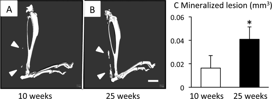
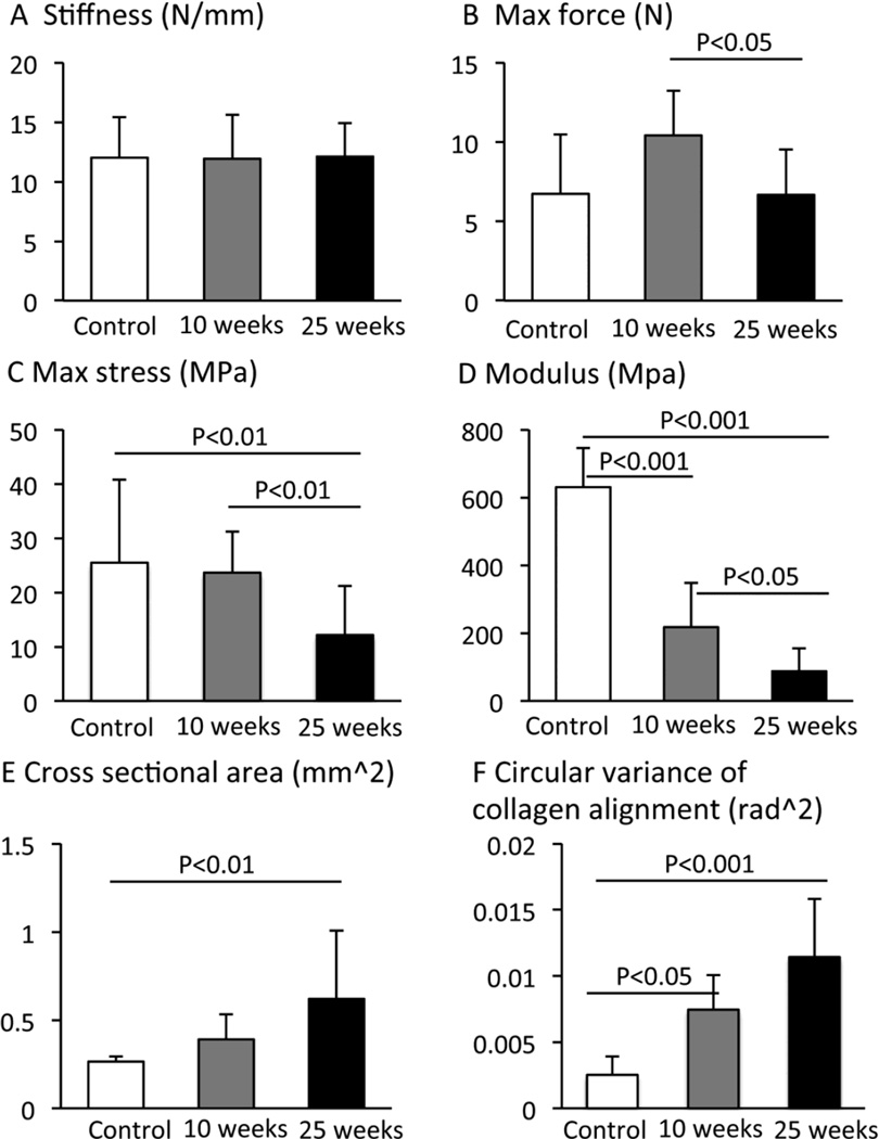
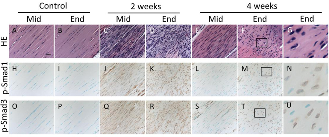
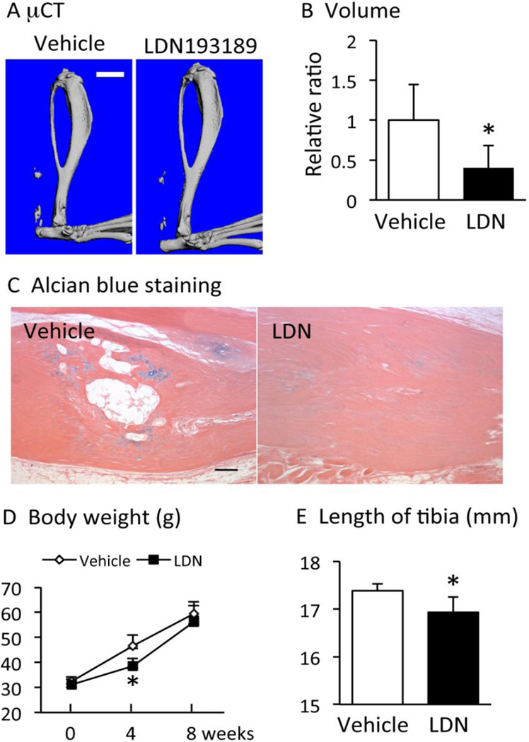
Ectopic tendon mineralization can develop following tendon rupture or trauma surgery. The pathogenesis of ectopic tendon mineralization and its clinical impact have not been fully elucidated yet. In this study, we utilized a mouse Achilles tendon injury model to determine whether ectopic tendon mineralization alters the biomechanical properties of the tendon and whether BMP signaling is involved in this condition. A complete transverse incision was made at the midpoint of the right Achilles tendon in 8-week-old CD1 mice and the gap was left open. Ectopic cartilaginous mass formation was found in the injured tendon by 4weeks post-surgery and ectopic mineralization was detected at 8 to 10weeks post-surgery. Ectopic mineralization grew over time and volume of the mineralized materials of 25-weeks samples was about 2.5 fold bigger than that of 10-weeks samples, indicating that injury-induced ectopic tendon mineralization is progressive. In vitro mechanical testing showed that max force, max stress and mid-substance modulus in the 25-weeks samples were significantly lower than the 10-weeks samples. We observed substantial increases in expression of bone morphogenetic protein family genes in injured tendons 1week post-surgery. Immunohistochemical analysis showed that phosphorylation of both Smad1 and Smad3 was highly increased in injured tendons as early as 1week post-injury and remained high in ectopic chondrogenic lesions 4-weeks post-injury. Treatment with the BMP receptor kinase inhibitor (LDN193189) significantly inhibited injury-induced tendon mineralization. These findings indicate that injury-induced ectopic tendon mineralization is progressive, involves BMP signaling and associated with deterioration of tendon biomechanical properties.
Keywords: Achilles tendon; BMP; Biomechanics; Ectopic mineralization; Injury; Smad.
Copyright © 2016 International Society of Matrix Biology. Published by Elsevier B.V. All rights reserved.
Figures
Similar articles
-
Tendon mineralization is accelerated bilaterally and creep of contralateral tendons is increased after unilateral needle injury of murine achilles tendons.J Orthop Res. 2013 Oct;31(10):1520-8. doi: 10.1002/jor.22404. Epub 2013 Jun 10. J Orthop Res. 2013. PMID: 23754538
-
BMP-14 gene therapy increases tendon tensile strength in a rat model of Achilles tendon injury.J Bone Joint Surg Am. 2007 Jun;89(6):1315-20. doi: 10.2106/JBJS.F.00257. J Bone Joint Surg Am. 2007. PMID: 17545436
-
Different mechanisms activated by mild versus strong loading in rat Achilles tendon healing.PLoS One. 2018 Jul 25;13(7):e0201211. doi: 10.1371/journal.pone.0201211. eCollection 2018. PLoS One. 2018. PMID: 30044869 Free PMC article.
-
Liquid Poly-N-acetyl Glucosamine (sNAG) Improves Achilles Tendon Healing in a Rat Model.Ann Biomed Eng. 2021 Feb;49(2):515-522. doi: 10.1007/s10439-020-02711-w. Epub 2021 Jan 6. Ann Biomed Eng. 2021. PMID: 33409852 Free PMC article. Review.
-
The Correlation of Dynamic Magnetic Resonance Imaging Evaluation With Histological, Biochemical, and Biomechanical Properties in Healing Progress After Achilles Tendon Injury: A Review.J Magn Reson Imaging. 2024 Oct;60(4):1243-1258. doi: 10.1002/jmri.29142. Epub 2023 Nov 22. J Magn Reson Imaging. 2024. PMID: 37991165 Review.
Cited by
-
Interplay of Forces and the Immune Response for Functional Tendon Regeneration.Front Cell Dev Biol. 2021 Jun 4;9:657621. doi: 10.3389/fcell.2021.657621. eCollection 2021. Front Cell Dev Biol. 2021. PMID: 34150755 Free PMC article. Review.
-
Profibrotic mediators in tendon disease: a systematic review.Arthritis Res Ther. 2016 Nov 18;18(1):269. doi: 10.1186/s13075-016-1165-0. Arthritis Res Ther. 2016. PMID: 27863509 Free PMC article.
-
Hedgehog signaling underlying tendon and enthesis development and pathology.Matrix Biol. 2022 Jan;105:87-103. doi: 10.1016/j.matbio.2021.12.001. Epub 2021 Dec 24. Matrix Biol. 2022. PMID: 34954379 Free PMC article. Review.
-
Bioinformatics analysis and experimental validation of key genes associated with lumbar disc degeneration and biomechanics.Heliyon. 2024 Feb 28;10(5):e27016. doi: 10.1016/j.heliyon.2024.e27016. eCollection 2024 Mar 15. Heliyon. 2024. PMID: 38463775 Free PMC article.
-
Treatment with Human Amniotic Suspension Allograft Improves Tendon Healing in a Rat Model of Collagenase-Induced Tendinopathy.Cells. 2019 Nov 8;8(11):1411. doi: 10.3390/cells8111411. Cells. 2019. PMID: 31717431 Free PMC article.
References
-
- Richards PJ, Braid JC, Carmont MR, Maffulli N. Achilles tendon ossification: pathology, imaging and aetiology. Disabil Rehabil. 2008;30(20–22):1651–1665. - PubMed
-
- Oliva F, Via AG, Maffulli N. Calcific tendinopathy of the rotator cuff tendons. Sports Med Arthrosc. 2011;19(3):237–243. - PubMed
-
- Kraus R, Stahl JP, Meyer C, Pavlidis T, Alt V, Horas U, Schnettler R. Frequency and effects of intratendinous and peritendinous calcifications after open Achilles tendon repair. Foot Ankle Int. 2004;25(11):827–832. - PubMed
Publication types
MeSH terms
Substances
Grants and funding
LinkOut - more resources
Full Text Sources
Other Literature Sources
Molecular Biology Databases
Miscellaneous
