Review
. 2016 Mar;73(5):1021-37.
doi: 10.1007/s00018-015-2107-8. Epub 2016 Jan 29.

Thymic stromal cell subsets for T cell development

Affiliations
Review

Thymic stromal cell subsets for T cell development

Takeshi Nitta et al. Cell Mol Life Sci. 2016 Mar.

Abstract

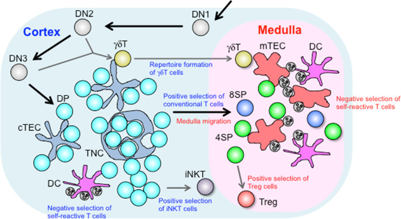
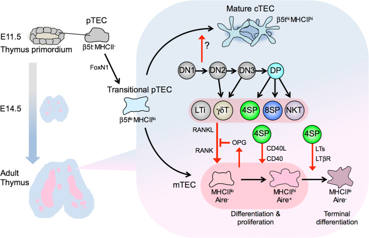
The thymus provides a specialized microenvironment in which a variety of stromal cells of both hematopoietic and non-hematopoietic origin regulate development and repertoire selection of T cells. Recent studies have been unraveling the inter- and intracellular signals and transcriptional networks for spatiotemporal regulation of development of thymic stromal cells, mainly thymic epithelial cells (TECs), and the molecular mechanisms of how different TEC subsets control T cell development and selection. TECs are classified into two functionally different subsets: cortical TECs (cTECs) and medullary TECs (mTECs). cTECs induce positive selection of diverse and functionally distinct T cells by virtue of unique antigen-processing systems, while mTECs are essential for establishing T cell tolerance via ectopic expression of peripheral tissue-restricted antigens and cooperation with dendritic cells. In addition to reviewing the role of the thymic stroma in conventional T cell development, we will discuss recently discovered novel functions of TECs in the development of unconventional T cells, such as natural killer T cells and γδT cells.

Keywords: Repertoire selection; T cell; Thymic epithelial cell; Thymus; cTEC; mTEC.


Figures

Fig. 1
Fig. 1
Roles of thymic stromal cells in T cell development. Developing DN thymocytes migrate from the cortico-medullary junction toward the subcapsular region, and then back toward the medulla upon development to DP thymocytes. In the cortex, DP thymocytes begin to express TCR on their surface and are selected upon interaction with pMHC complexes displayed in the microenvironment. cTECs produce a unique set of MHC-bound peptides that are essential for inducing positive selection, while DCs critically contribute to induction of negative selection. Intimate interaction between DP thymocytes and cTECs results in formation of TNCs, which facilitate prolonged survival of inner DP thymocytes and secondary TCRα recombination. Positively selected cells migrate into the medulla in response to chemokines produced by mTECs. γδT cells diverge from the αβT cell lineage at the DN stage, and their repertoire formation is regulated by cTECs through unknown mechanisms. The cortical microenvironment is also important for positive selection of iNKT cells that depends on cell–cell interaction among DP thymocytes. In the medulla, SP thymocytes are screened for self-reactivity. SP thymocytes reactive to TRAs presented by mTECs or DCs are deleted by negative selection or induced to differentiate into Foxp3+ Treg cells
Fig. 2
Fig. 2
Development of thymic epithelial cells is induced by various subsets of thymocytes. cTECs and mTECs arise from common progenitor TECs (pTECs) in endodermal epithelium from the third pharyngeal pouch. pTECs differentiate into ‘transitional’ pTECs that express cTEC-associated genes such as β5t and IL-7. This process is critically regulated by the transcription factor FoxN1 but independent of lymphocytes. Thymocyte development beyond DN1 induces maturation of cTECs expressing high levels of β5t. The development of mTECs is triggered in embryonic thymus by LTi cells and γδT cells that express RANKL. In postnatal thymus, SP thymocytes and NKT cells express RANKL to promote the differentiation and proliferation of Aire-expressing mTECs. RANKL-stimulated mTECs produce OPG to self-tune their development. CD40L expressed in CD4SP thymocytes cooperates with RANKL to promote mTEC development. CD4SP thymocytes also express LTs, which induce terminal differentiation of mTECs

References

    1. Von Boehmer H. Selection of the T-cell repertoire: receptor-controlled checkpoints in T-cell development. Adv Immunol. 2004;84(84):201–238. doi: 10.1016/S0065-2776(04)84006-9. - DOI - PubMed
    1. Hogquist KA, Jameson SC. The self-obsession of T cells: how TCR signaling thresholds affect fate ‘decisions’ and effector function. Nat Immunol. 2014;15(9):815–823. doi: 10.1038/ni.2938. - DOI - PMC - PubMed
    1. Miller JF. Immunological function of thymus. Lancet. 1961;2(720):748–749. doi: 10.1016/S0140-6736(61)90693-6. - DOI - PubMed
    1. Boyd RL, Tucek CL, Godfrey DI, Izon DJ, Wilson TJ, Davidson NJ, Bean AGD, Ladyman HM, Ritter MA, Hugo P. The thymic microenvironment. Immunol Today. 1993;14(9):445–459. doi: 10.1016/0167-5699(93)90248-J. - DOI - PubMed
    1. Gray DH, Ueno T, Chidgey AP, Malin M, Goldberg GL, Takahama Y, Boyd RL. Controlling the thymic microenvironment. Curr Opin Immunol. 2005;17(2):137–143. doi: 10.1016/j.coi.2005.02.001. - DOI - PubMed

Publication types

LinkOut - more resources