. 2016 May 1;110(1):51-61.
doi: 10.1093/cvr/cvw024. Epub 2016 Jan 29.

Temporal neutrophil polarization following myocardial infarction

Affiliations

Temporal neutrophil polarization following myocardial infarction

Yonggang Ma et al. Cardiovasc Res. .

Abstract

Aims: Although macrophage phenotypes have been well studied in the myocardial infarction (MI) setting, this study investigated temporal neutrophil polarization and activation mechanisms.

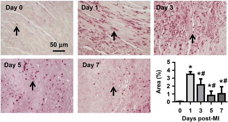
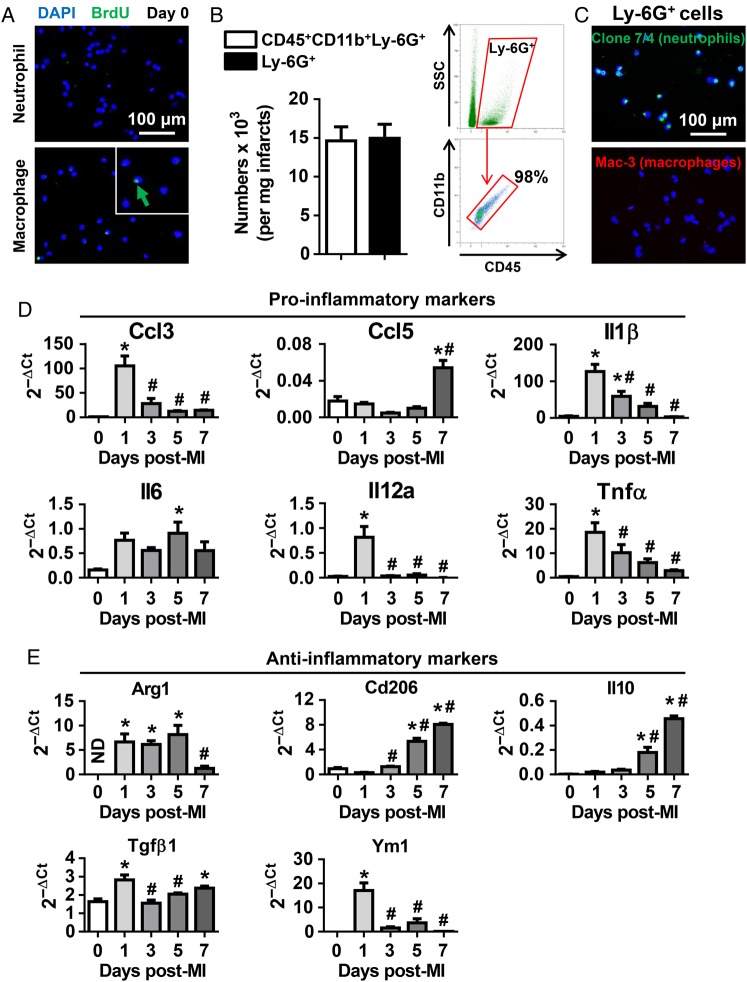
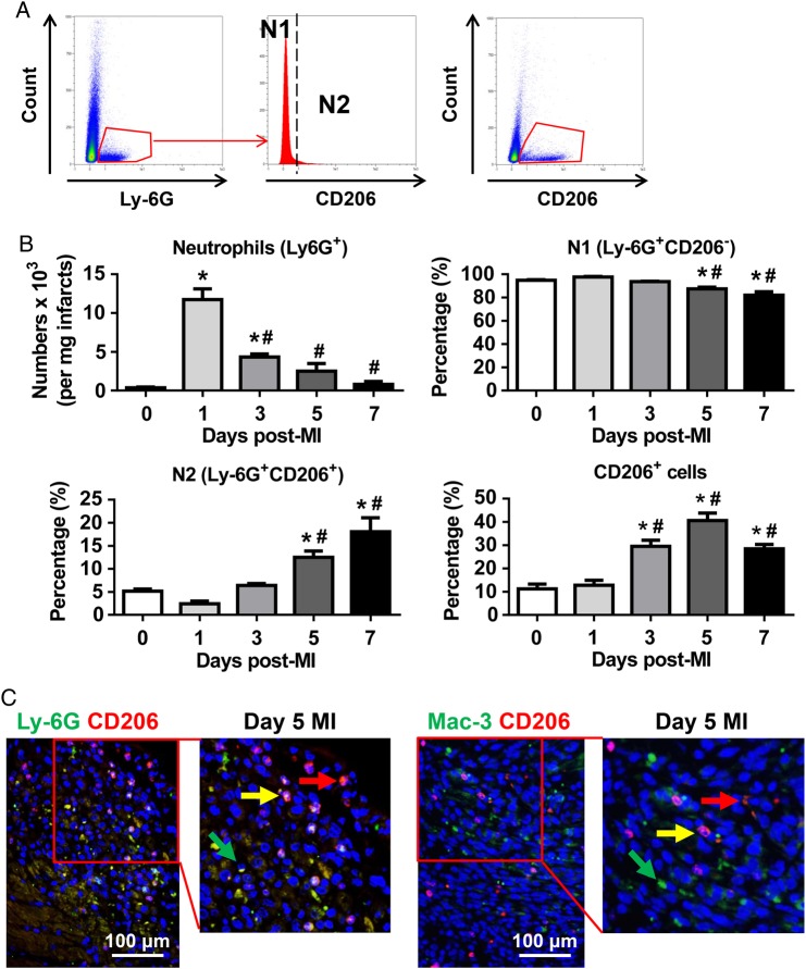
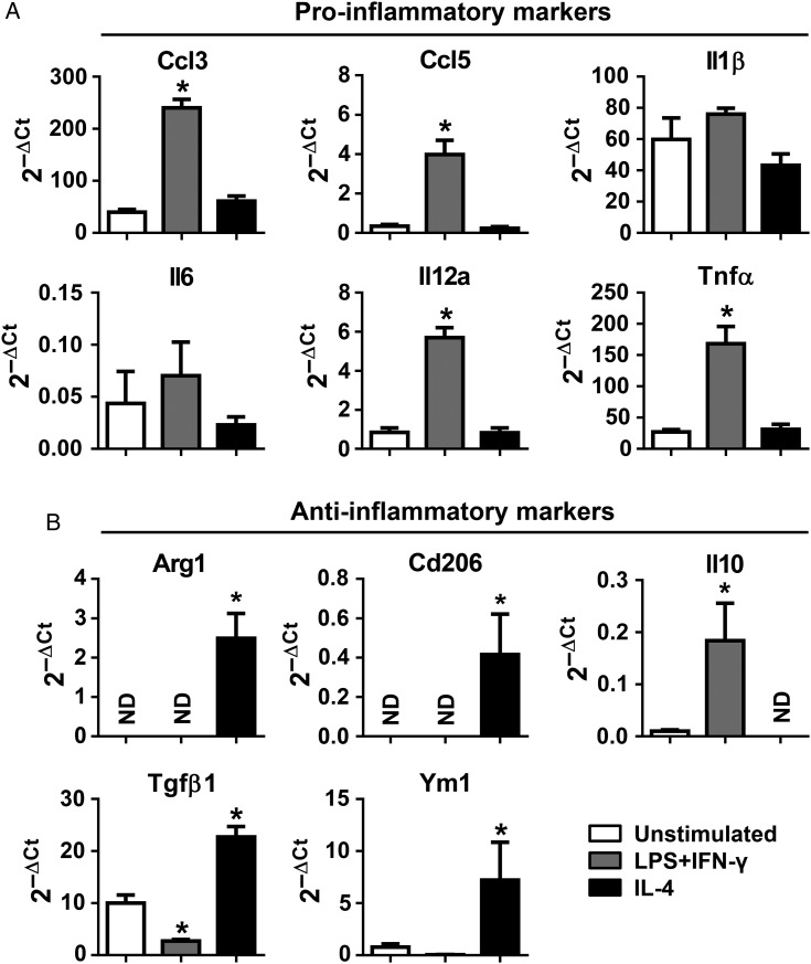
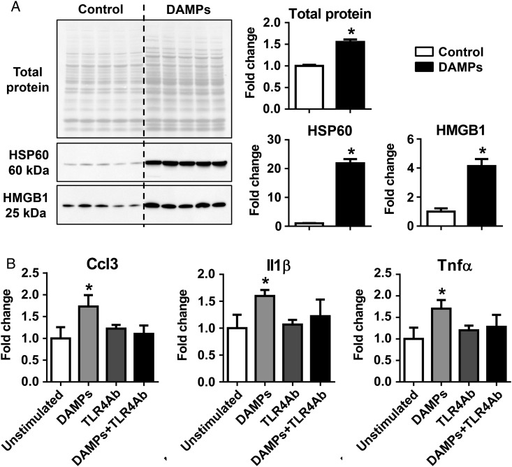
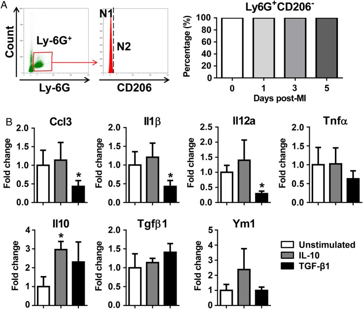
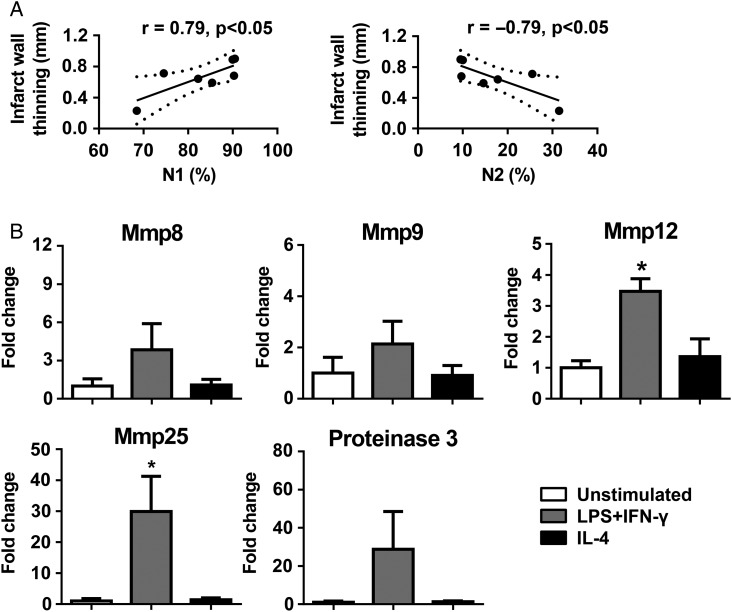
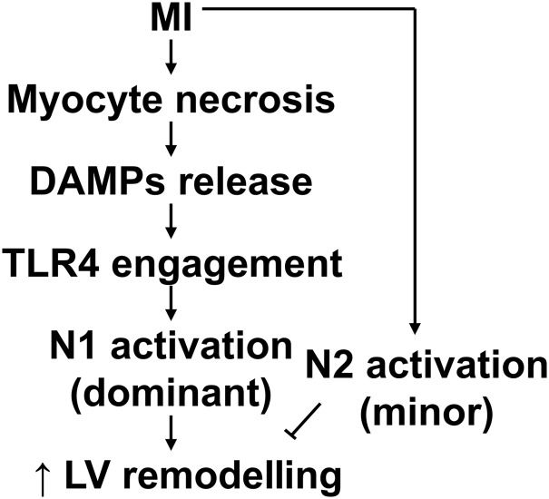
Methods and results: Neutrophils isolated from the infarcted left ventricle (LV) of mice showed high expression of proinflammatory markers at Day 1 and anti-inflammatory markers at Days 5 and 7 post-MI, indicating distinct neutrophil phenotypes along the post-MI time continuum. Flow cytometry analysis revealed that although proinflammatory N1 neutrophils were always predominant (>80% of total neutrophils at each time point), the percentage of N2 neutrophils increased post-MI from 2.4 ± 0.6% at Day 1 to 18.1 ± 3.0% at Day 7. In vitro, peripheral blood neutrophils were polarized to proinflammatory N1 by lipopolysaccharide and interferon-γ or anti-inflammatory N2 by interleukin-4, indicating high plasticity potential. The in vivo post-MI relevant LV damage-associated molecular patterns (DAMPs) polarized neutrophils to a proinflammatory N1 phenotype by activating toll-like receptor-4. Transforming growth factor-β1 inhibited proinflammatory production in neutrophils. N1 neutrophils positively correlated with infarct wall thinning at Day 7 post-MI, possibly due to high production of matrix metalloproteinases-12 and -25.

Conclusion: This study is the first to identify the existence of N1 and N2 neutrophils in the infarct region and reveals that N1 polarization could be mediated by DAMPs.

Keywords: DAMPs; Inflammation; Myocardial infarction; Neutrophil polarization; Proteomics.


Figures

Figure 1
Figure 1
Temporal neutrophil infiltration. Time course of neutrophil infiltration post-MI. At Day 0 (no MI), very few neutrophils were observed in the LV. After MI, neutrophil infiltration peaked at Days 1–3, declined at Day 5, and was low by Day 7. Arrows indicate neutrophils. n = 8–16 per group. One-way ANOVA was used.
Figure 2
Figure 2
The expression of proinflammatory and anti-inflammatory markers in neutrophils. (A) Representative images demonstrated that cardiac resident neutrophils (Day 0) did not proliferate, as evidenced by BrdU-negative staining. Macrophages served as positive controls. (B) The numbers of CD45+CD11b+Ly-6G+ cells and Ly-6G+ cells at Day 1 post-MI evaluated by flow cytometry. n = 6 per group. (C) Ly6G+ cells were positive for the neutrophil clone 7/4, but negative for the macrophage marker Mac-3. Representative images of four biological samples. (D and E) Neutrophils expressed high levels of proinflammatory markers at Day 1 and anti-inflammatory markers at Days 5 and 7 post-MI, as assessed by quantitative RT–PCR. ND, not determined (value below detection). We assigned ND as zero when performing statistical analysis. Due to the small number of neutrophils in Day 0 LVs, we pooled three to four LVs per sample. n = 4–6 samples per group. *P < 0.05 vs. Day 0 and #P < 0.05 vs. Day 1 post-MI. One-way ANOVA was used.
Figure 3
Figure 3
Temporal neutrophil polarization in the infarcted LVs. (A) Representative flow cytometry plots illustrate gating strategy to identify neutrophil N1 and N2 phenotypes and CD206+ cells. (B) Although proinflammatory N1 was the predominant neutrophil in the infarcted LVs at all times examined, the percentage of anti-inflammatory N2 neutrophils increased over the course of MI. n = 6–12 per group. *P < 0.05 vs. Day 0 and #P < 0.05 vs. Day 1 post-MI. One-way ANOVA was used. (C) Dual immunofluorescence images revealed the presence of Ly-6G+CD206 N1 (green arrow, left panel), Ly-6G+CD206+ N2 (yellow arrow, left panel), Mac-3+CD206 M1 (green arrow, right panel), Mac-3+CD206+ M2 (yellow arrow, right panel), and CD206+ cells (red arrow) in day 5 infarcted LVs. Representative images of three to six biological samples.
Figure 4
Figure 4
LPS and IFN-γ polarized neutrophils to a proinflammatory N1 phenotype, whereas IL4 polarized neutrophils to an anti-inflammatory N2 phenotype. (A and B) LPS (1 μg/mL) and IFN-γ (20 ng/mL) up-regulated the expression of proinflammatory markers; on the contrary, IL-4 (20 ng/mL) treatment up-regulated the expression of anti-inflammatory markers in neutrophils. ND, not determined (value below detection). We assigned ND as zero when performing statistical analysis. n = 5 mice per sample. n = 5–6 samples per group. *P < 0.05 vs. unstimulated. One-way ANOVA was used.
Figure 5
Figure 5
DAMPs activated N1 neutrophil polarization in vitro via TLR4. (A) LV tissues that underwent freeze–thaw cycles (the DAMPs group) contained significantly higher total protein, HSP60, and HMGB1, compared with controls, as evaluated by immunoblotting. n = 5 LVs per group. The same volume of all samples was loaded (2 µL). (B) DAMPs stimulated the production of proinflammatory markers Ccl3, Il1β, and Tnfα, which was abolished by the anti-TLR4 neutralizing antibody. The data were expressed as fold change (normalized to control or unstimulated group). n = 3–6 mice per sample and n = 4–9 samples per group; *P < 0.05 vs. controls. One-way ANOVA was used.
Figure 6
Figure 6
CD206+ N2 neutrophils were activated in the post-MI LV, but not in circulation. (A) Circulating neutrophils at different time points post-MI were CD206. n = 6–8 per group. (B) IL-10 (50 ng/mL) up-regulated the expression of the anti-inflammatory marker Il10; TGF-β1 (10 ng/mL) inhibited the expression of proinflammatory markers (Ccl3, Il1β, and Il12a) in neutrophils. The data were expressed as fold change (normalized to the unstimulated group). n = 4 mice per sample and n = 4 samples per group. *P < 0.05 vs. unstimulated. One-way ANOVA was used.
Figure 7
Figure 7
N1 neutrophils were a source of LV wall thinning due to high levels of Mmp12 and Mmp25. (A) At day 7 post-MI, N1 positively and N2 negatively correlated with infarct wall thinning. Infarct wall thinning = (days 0–7) LV posterior wall thickness in systole. n = 7 per group. Pearson's correlation analysis was used. (B) N1 neutrophils stimulated by LPS + IFN-γ expressed higher levels of Mmp12 and Mmp25, compared with unstimulated cells. The data were expressed as fold change (normalized to the unstimulated group). n = 4 per group. *P < 0.05 vs. unstimulated. One-way ANOVA was used.
Figure 8
Figure 8
Mechanisms of neutrophil N1 polarization post-MI. Following MI, necrotic myocytes release DAMPs to polarize neutrophils to a proinflammatory N1 phenotype by activating TLR4. N1 may exacerbate and N2 may attenuate adverse LV remodelling.

References

    1. Ghasemi O, Ma Y, Lindsey ML, Jin YF. Using systems biology approaches to understand cardiac inflammation and extracellular matrix remodeling in the setting of myocardial infarction. Wiley Interdiscip Rev Syst Biol Med 2014;6:77–91. - PMC - PubMed
    1. Ma Y, Yabluchanskiy A, Lindsey ML. Neutrophil roles in left ventricular remodeling following myocardial infarction. Fibrogenesis Tissue Repair 2013;6:11. - PMC - PubMed
    1. Cui BB, Tan CY, Schorn C, Tang HH, Liu Y, Zhao Y. Neutrophil extracellular traps in sterile inflammation: the story after dying? Autoimmunity 2012;45:593–596. - PubMed
    1. Mantovani A, Cassatella MA, Costantini C, Jaillon S. Neutrophils in the activation and regulation of innate and adaptive immunity. Nat Rev Immunol 2011;11:519–531. - PubMed
    1. Romson JL, Hook BG, Kunkel SL, Abrams GD, Schork MA, Lucchesi BR. Reduction of the extent of ischemic myocardial injury by neutrophil depletion in the dog. Circulation 1983;67:1016–1023. - PubMed

Publication types