

Case Reports
doi: 10.1097/WAD.0000000000000130.
Heterozygous Chorein Deficiency in Probable Tau-negative Early-onset Alzheimer Disease
Affiliations
- PMID: 26825611
- PMCID: PMC5035148
- DOI: 10.1097/WAD.0000000000000130
Item in Clipboard
Case Reports
Heterozygous Chorein Deficiency in Probable Tau-negative Early-onset Alzheimer Disease
Alzheimer Dis Assoc Disord.
2016 Jul-Sep.
Abstract
Supplemental Digital Content is available in the text.
Conflict of interest statement
The authors declare no conflicts of interest.
Figures
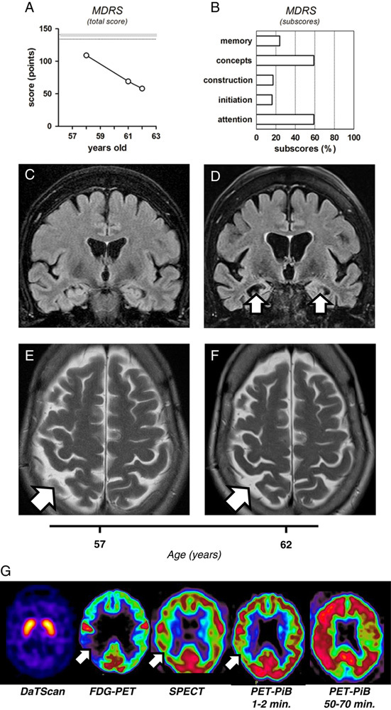
Neurocognitive and brain structural and metabolic assessment of the reported patient. A, Changes in the total score of Mattis dementia rating scale (MDRS), indicating progressive cognitive decline (109/144, 69/144, and 58/144, at the age of 58, 61, and 62, respectively). Shadowed range indicated on the panel represents a 25 to 75 percentile interval, previously established for the population younger than 70 years old. B, Specific MDRS subscores collected at the age of 62, and expressed as a percentage of the maximal possible value for each subscore. C-D, The coronal FLAIR imaging, demonstrating a clear progression of the hippocampal atrophy (white arrows) within 5 years of follow-up, from the age of 57 (C) to 62 (D). Note the right hemispheric dominance of the hippocampal atrophy and consecutive dilation of the temporal horns of the lateral ventricles. E-F, The axial T2 shows a mild parietal atrophy (white arrows), again with right hemispheric predominance, which only slowly progressed between the age to 57 (E) and 62 (F). Note the absence of microvascular white matter lesions. G, Metabolic neuroimaging. DaTScan imaging shows normal uptake of the tracer in the caudate and putamen. FDG-PET indicates global decrease in cerebral metabolism, particularly severe in the right temporo-parietal region (white arrow). SPECT reveals generalized hypoperfusion, slightly predominant on the right side (white arrow). Positron-emission tomography with Pittsburgh compound-B (PET-PiB) shows decreased tracer activity after 1 to 2 minutes, especially in the right temporo-parietal region (white arrow), and massive and widespread retention of PiB after 50 to 70 minutes, again with right predominance.

Chorein deficiency and its impact on amyloid processing. A, Family tree, with the proband indicated by black arrow. The proband and 2 of 3 of her children presented with neuropsychiatric disorders (black symbols; clinical details in the text). Clinical profile of other family members remained undetermined (white symbols). B, Illustration of the results of genetic analysis. Deleted exons in chorein are marked with gray asterisk. The alternative splicing site is marked with black asterisk, and the exon 69 (skipped in the canonical form of chorein) is shadowed. C–F, In vitro chorein knockdown system. The neuronal SH-SY5Y cells were transfected with specific, chorein-targeting siRNA (white bars) or negative control siRNA (black bars). Expression of chorein (C), as well as APP, PSEN1, PSEN2, and SORL1 (E) was assessed by quantitative PCR and expressed as a percentage of its respective negative control; chorein knockdown had no impact on the viability/proliferation of SH-SY5Y cells (D); concentration of Aβ40, but not Aβ42, was significantly decreased in supernatants from SH-SY5Y cells upon chorein knockdown (F); concentration of Aβ40 was low in the cerebrospinal fluid of the proband, as compared with mean reference values previously reported for healthy controls (black arrow and dashed line) and demented patients carrying PSEN1 pathogenic mutations (gray arrow and dashed line) (G). For quantitative PCR analysis 7 (n=7; C) or 4 (n=4; E) independent experiments were performed. For cell counting (D) and measurement of Aβ concentrations (F), 5 independent experiments were performed (n=5). All of the values in the panels (C–E) are presented as percentages of the respective control groups (means±SEM). The significance of the differences (chorein knockdown vs. control) was tested by the 2-tailed Mann-Whitney nonparametric test (E), or by the 2-tailed paired t test for the sets of data following normal distribution (C, D, F; normal distribution confirmed by Kolmogorov-Smirnov test). The difference between the means were considered significant if P<0.05. NS indicates statistically not significant; **P<0.01.
Comment in
-
Heterozygous Deletion of Chorein Exons 70-73 and GNA14 Exons 3-7 in a Brazilian Patient Presenting With Probable Tau-Negative Early-Onset Alzheimer Disease.Alzheimer Dis Assoc Disord. 2017 Jan-Mar;31(1):82-85. doi: 10.1097/WAD.0000000000000185. Alzheimer Dis Assoc Disord. 2017. PMID: 28079535 No abstract available.
-
Chorein Deficiency and Alzheimer Disease: An Intriguing, Yet Premature Speculation.Alzheimer Dis Assoc Disord. 2017 Jan-Mar;31(1):80-81. doi: 10.1097/WAD.0000000000000186. Alzheimer Dis Assoc Disord. 2017. PMID: 28121633 Free PMC article. No abstract available.
References
-
- Klünemann HH, Fronhöfer W, Werner-Füchtenbusch D, et al. Characterization of the kindred of Alois Alzheimer’s patient with plaque-only dementia. Alzheimer Dis Assoc Disord. 2006;20:291–294. - PubMed
-
- Rampoldi L, Dobson-Stone C, Rubio JP, et al. A conserved sorting-associated protein is mutant in chorea-acanthocytosis. Nat Genet. 2001;28:119–120. - PubMed
-
- Hayashi T, Kishida M, Nishizawa Y, et al. Subcellular localization and putative role of VPS13A/chorein in dopaminergic neuronal cells. Biochem Biophys Res Commun. 2012;419:511–516. - PubMed
-
- Dobson-Stone C, Danek A, Rampoldi L, et al. Mutational spectrum of the CHAC gene in patients with chorea-acanthocytosis. Eur J Hum Genet. 2002;10:773–781. - PubMed
-
- Tomiyasu A, Nakamura M, Ichiba M, et al. Novel pathogenic mutations and copy number variations in the VPS13A gene in patients with chorea-acanthocytosis. Am J Med Genet B Neuropsychiatr Genet. 2011;156B:620–631. - PubMed
Publication types
MeSH terms
Substances
LinkOut - more resources
Full Text Sources
Other Literature Sources
Medical
