

Age-related changes in murine bladder structure and sensory innervation: a multiphoton microscopy quantitative analysis
- PMID: 26825637
- PMCID: PMC5005881
- DOI: 10.1007/s11357-016-9878-1
Age-related changes in murine bladder structure and sensory innervation: a multiphoton microscopy quantitative analysis
Abstract
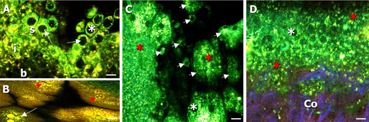
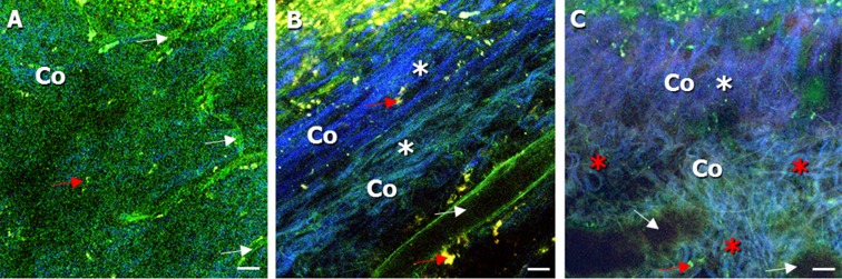
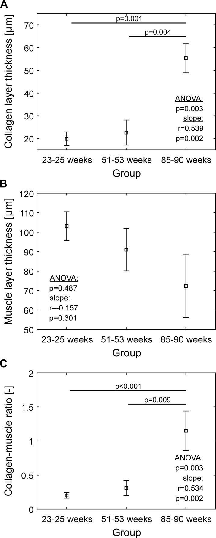
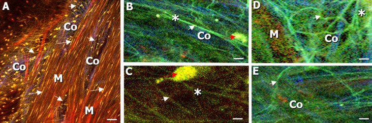
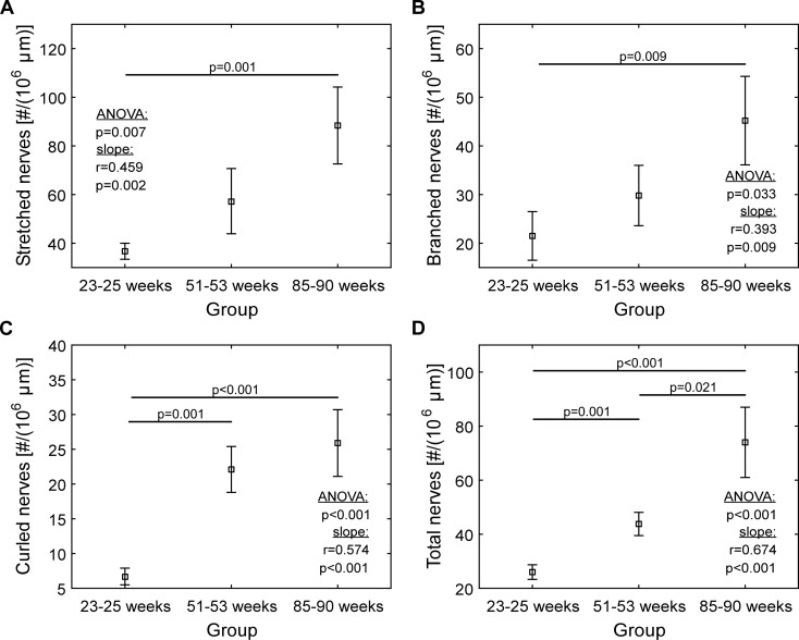
Our study aimed to examine and quantify age-related structural alterations in the healthy mouse bladder using ex vivo two-photon laser scanning microscopy (TPLSM). Freshly dissected bladders from 25-, 52-, and 85-week-old C57bl/6J mice were examined, and morphological analyses and quantification of cell layers and nerves were performed. The numbers of stretched, curled, branched, and total number of nerves in volume units of the stained muscle layer were quantified. We observed differences in the bladder wall architecture and innervation with age. Especially in 85-week-old mice, age-related changes were found, including detachment of urothelial cells and an increase in connective tissue, intermingled with the smooth muscle fibers in the muscle layer (collagen-smooth muscle ratio of 1.15 ± 0.29). In 25- and 52-week-old mice, the collagen-smooth muscle ratios were 0.20 ± 0.04 and 0.31 ± 0.11, respectively, and a clear separation of collagen and muscle was observed. The overall number of nerves and the number of curled nerves were significantly higher in the 85-week-old mice (74.0 ± 13.0 and 25.9 ± 4.8, respectively), when comparing to 25-week-old mice (26.0 ± 2.7 and 6.7 ± 1.2, respectively) and 52-week-old mice (43.8 ± 4.3 and 22.1 ± 3.3, respectively). Significant age-related alterations in bladder morphology and innervation were found, when comparing freshly dissected bladder tissue from 25-, 52-, and 85-week-old mice. The higher number of curled nerves might be an indication of an increased neurotransmitter release, resulting in a higher nerve activity, with a part of the nerves being possibly mechanically impaired. This study shows that two-photon laser scanning microscopy of healthy aging male mice is a useful method to investigate and quantify the age-related changes in the bladder wall.
Keywords: Aging; Mice; Multiphoton microscopy; Sensory innervation; Urinary bladder.
Conflict of interest statement
Compliance with ethical standards Experimental protocols were approved by the animal ethics committee of Maastricht University, and were carried out according to institutional guidelines, and reported in accordance with the ARRIVE guidelines.
Figures

Similar articles
-
Computer-assisted three-dimensional tracking of sensory innervation in the murine bladder mucosa with two-photon microscopy.J Chem Neuroanat. 2017 Nov;85:43-49. doi: 10.1016/j.jchemneu.2017.06.006. Epub 2017 Jun 28. J Chem Neuroanat. 2017. PMID: 28668578
-
Changes in bladder innervation in a mouse model of Alzheimer's disease.J Chem Neuroanat. 2010 May;39(3):204-10. doi: 10.1016/j.jchemneu.2009.12.001. Epub 2009 Dec 16. J Chem Neuroanat. 2010. PMID: 20025962
-
Quantitative multiphoton microscopy of murine urinary bladder morphology during in situ uniaxial loading.Acta Biomater. 2017 Dec;64:59-66. doi: 10.1016/j.actbio.2017.09.029. Epub 2017 Sep 22. Acta Biomater. 2017. PMID: 28951123
-
Control of bladder function by peripheral nerves: avenues for novel drug targets.Urology. 2004 Mar;63(3 Suppl 1):24-31. doi: 10.1016/j.urology.2003.10.031. Urology. 2004. PMID: 15013649 Review.
-
Innervation of bladder and bowel.Ciba Found Symp. 1990;151:2-18; discussion 18-26. doi: 10.1002/9780470513941.ch2. Ciba Found Symp. 1990. PMID: 1977565 Review.
Cited by
-
A Novel in situ Approach to Studying Detrusor Smooth Muscle Cells in Mice.Sci Rep. 2020 Feb 14;10(1):2685. doi: 10.1038/s41598-020-59337-0. Sci Rep. 2020. PMID: 32060298 Free PMC article.
-
Effect of filling rate on cystometric parameters in young and middle aged mice.Bladder (San Franc). 2017;4(1):e28. doi: 10.14440/bladder.2017.88. Epub 2017 Feb 27. Bladder (San Franc). 2017. PMID: 28553656 Free PMC article.
-
Aging and urinary control: Alterations in the brain-bladder axis.Aging Cell. 2023 Dec;22(12):e13990. doi: 10.1111/acel.13990. Epub 2023 Sep 22. Aging Cell. 2023. PMID: 37740454 Free PMC article. Review.
-
Modeling the influence of acute changes in bladder elasticity on pressure and wall tension during filling.J Mech Behav Biomed Mater. 2017 Jul;71:192-200. doi: 10.1016/j.jmbbm.2017.02.020. Epub 2017 Feb 20. J Mech Behav Biomed Mater. 2017. PMID: 28343086 Free PMC article.
-
Age-Related Changes in Neuromodulatory Control of Bladder Micturition Contractions Originating in the Skin.Front Neurosci. 2018 Feb 27;12:117. doi: 10.3389/fnins.2018.00117. eCollection 2018. Front Neurosci. 2018. PMID: 29599702 Free PMC article.
References
-
- Al-Motabagani M. Age-related changes in the urinary bladder of the female albino rats. Int J Morphol. 2005;23(4):309–316.
Publication types
MeSH terms
LinkOut - more resources
Full Text Sources
Other Literature Sources
Medical