. 2016 Feb 16;44(2):259-73.
doi: 10.1016/j.immuni.2015.11.017. Epub 2016 Jan 26.

T Follicular Helper Cell Plasticity Shapes Pathogenic T Helper 2 Cell-Mediated Immunity to Inhaled House Dust Mite

Affiliations

T Follicular Helper Cell Plasticity Shapes Pathogenic T Helper 2 Cell-Mediated Immunity to Inhaled House Dust Mite

André Ballesteros-Tato et al. Immunity. .

Abstract

Exposure to environmental antigens, such as house dust mite (HDM), often leads to T helper 2 (Th2) cell-driven allergic responses. However, the mechanisms underlying the development of these responses are incompletely understood. We found that the initial exposure to HDM did not lead to Th2 cell development but instead promoted the formation of interleukin-4 (IL-4)-committed T follicular helper (Tfh) cells. Following challenge exposure to HDM, Tfh cells differentiated into IL-4 and IL-13 double-producing Th2 cells that accumulated in the lung and recruited eosinophils. B cells were required to expand IL-4-committed Tfh cells during the sensitization phase, but did not directly contribute to disease. Impairment of Tfh cell responses during the sensitization phase or Tfh cell depletion prevented Th2 cell-mediated responses following challenge. Thus, our data demonstrate that Tfh cells are precursors of HDM-specific Th2 cells and reveal an unexpected role of B cells and Tfh cells in the pathogenesis of allergic asthma.


Figures

Figure 1
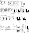
Figure 1. B cells control HDM-induced pulmonary Th2 cell responses
(A-E) B6.4get and μMT.4get mice were i.n treated with 25μg of HDM for 4 consecutive days starting on day 1. On day 15, mice were i.n challenged with 25μg of HDM daily for 4 days and analyzed on day 20 by flow cytometry (A). As a control, naïve B6.4get mice were also analyzed. The frequency (B and D) and number (C and E) of IL-4-expressing (EGFP+) CD4+ T cells from the lungs (B and C) and mLN (D and E) are shown. (F-G) The number of eosinophils in the lungs (F) and BAL (G) of naive and HDM-challenged B6 and μMT mice are shown. (H) Arterial oxygen saturation (SpO2) of hemoglobin, measured with pulse oximeter, 2 hours after final challenge. (I-K) B6 and μMT mice were i.n treated with 25μg of HDM for 4 consecutive days starting on day 1. Some μMT mice received 50×106 naïve B cells i.v. on day 0, or on day 14. On day 15, all mice were i.n challenged with 25μg of HDM daily for 4 consecutive days. Mice were analyzed on day 20. The frequency (I) and number (J) of IL-4+ CD4+ T cells in the lungs were determined by intracellular staining after restimulation for 4 h with anti-CD3 and BFA. (K) The number of eosinophils in the lungs are shown. (L-M) B6 and μMT mice were i.n treated with 25μg of HDM for 4 consecutive days starting on day 1. Six days later, CD4+ T cells were purified from the mLN and 2 × 10⁶ of purified CD4+ T cells from B6 or μMT donor mice were transferred into day 6 HDM-sensitized B6.CD45.1+ recipient mice. Recipient mice were i.n challenged on day 15 with 25μg of HDM daily for 4 consecutive days and analyzed on day 20 (L). The frequency (M) and number (N) of donor IL-4+ CD4+ T cells in the lungs of recipient mice were determined by intracellular staining after restimulation with anti-CD3+ BFA for 4 h. *P < 0.01 vs. HDM-treated B6. (unpaired Student’s t test). Data are representative of three independent experiments (mean and S.D. of 4-5 mice per group). See also Figure S1.
Figure 2
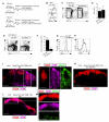
Figure 2. Ag presentation by B cells is necessary to prime of IL-4-commited CD4+ T cell responses to HDM
(A-B) IL-4 reporter B6.4get and μMT.4get mice were i.n treated with 25μg of HDM or PBS for 4 consecutive days starting on day 1. The frequency (A) and number (B) of IL-4-expressing (EGFP+) CD4+ T cells in the mLN of treated mice were calculated by flow cytometry on day 6. (C-H) μMT.4get mice were irradiated and reconstituted with an 80:20 mixture of BM from μMT.4get and B6 donors (B-WT), or with an 80:20 mixture of BM from μMT.4get and MHC II deficient donors (B-MHC II−/−). Eight weeks later, reconstituted-chimeric mice were i.n treated with 25μg of HDM of PBS for 4 consecutive days starting on day 1 and mice were analyzed at day 6. The frequency (D) and number (E) of B cells in the mLN were calculated by flow cytometry. (F) Expression of MHC II on B cells. Frequency (G) and number (H) of IL-4-expressing (EGFP+) CD4+ T cells in the mLN. *P < 0.001 vs. HDM-treated B6 (unpaired Student’s t test). Data are representative of three independent experiments (mean and S.D. of 4-5 mice per group).
Figure 3
Figure 3. Ag presentation by DC cells is necessary for the priming of specific CD4+ T cells after allergic sensitization
(A-H) B6 mice were irradiated and reconstituted with an 80:20 mixture of BM from CD11c-DTR and B6 donors (DC-WT) or from CD11c-DTR and MHC II−/− donors (DCMHC II−/−) (A). Eight weeks later, 25×103 naïve OTII.CD45.1+ CD4+ T cells were transferred into reconstituted chimeric mice on day 0. Recipient mice were treated with HDM+OVA (from day 1 to 4), injected with PBS or DT at day 0 and 3, and analyzed on day 6. The expression of MHC II on DC (B) and the frequency (C) and number (D) of DCs in the mLN are shown. Frequency (E) and number (F) of donor OTII T cells from the mLN. Expression of CD44 (G) and CD69 (H) in donor OTII T cells from the mLN. (I-M) B6 mice were i.n treated with 25μg of Alexa Fluor 647-labeled HDM. Cryosections from the mLN were stained with anti-B220 (red) and CD11c (green) and analyzed by fluorescent microscopy on days 1 (I-K) and 3 (L-M). The regions within the box in panels (I and L) were enlarged in panel (J and M) respectively. In the same experiment, CD11c-DTR mice were injected i.p. with 60 ng DT and i.n treated with 25μg of Alexa Fluor 647-labeled HDM 24 h later. Cryosections from the mLN one day 1 after HDM inoculation are shown (K). Scale bars, 400 μm. *P < 0.005 (unpaired Student’s t test). Data are representative of three independent experiments (mean and S.D. of 4-5 mice per group).
Figure 4
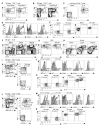
Figure 4. HDM sensitization induces IL-4-expressing Tfh cells but not effector Th2 cells
(A-E) B6.4get mice were i.n treated with 25μg of HDM of PBS for 4 consecutive days starting on day 1 and analyzed by flow cytometry on day 6. The frequency of IL-4-expressing (EGFP+) on the CD4+ T cells from the lungs (A) and mLNs (B) are shown. Expression of PD-1 and CXCR5 (C), BCL-6, GL7, CD25 and ICOS (D) and intracellular IL-2 and IL-21 (E) on naïve (CD44loEGFP) and antigen-experienced (CD44hiEGFP+) CD4+ T cells in the mLN of HDM-treated mice are shown at day 6. (F-H) B6 mice were i.n HDM sensitized on day 1 and analyzed on day 6 or HDM sensitized on day 1, challenged on day 15 and analyzed on day 20. The frequency of CD138+ antibody secreting cells (ASC) (F), IgE+CD138+ ACS (G) and PNA+FAS+ GC cells (H) in the mLN are shown. (I-N) B6.4get mice were i.n treated with 25μg of HDM for 4 consecutive days starting on day 1. On day 15, mice were i.n challenged with 25μg HDM daily for 4 consecutive days. Mice were sacrificed and analyzed by flow cytometry on day 20. The expression of PD-1 and CXCR5 (I), BCL-6, GL7, CD25 , ICOS and GATA-3 (J) and intracellular IL-2 and IL-21 (K) on naïve (CD44loEGFP) and antigen-experienced (CD44hiEGFP+) CD4+ T cells in the lung are shown. Expression of PD-1 and CXCR5 (L), BCL-6, GL7, CD25, ICOS and GATA-3 (M) and intracellular IL-2 and IL-21 (N) on naïve (CD44loEGFP) and antigen-experienced (CD44hiEGFP+) CD4+ T cells in the mLN. Data are representative of three independent experiments (mean and S.D. of 4-5 mice per group). See also Figure S2.
Figure 5
Figure 5. CD4 T cells activated after sensitization develop into effector Th2 cells after HDM challenge
(A-F) 25×103 naïve OTII.CD45.1+ CD4+ T cells were transferred into naive B6 and μMT mice. One day later, recipient mice were i.n treated with 25μg of HDM + 25μg of OVA (HDM+OVA), or PBS for 4 consecutive days. Mice were sacrificed and analyzed by flow cytometry 2 days after the last HDM inoculation. Frequency (A) and number (B) of donor OTII T cells from the mLN. Expression of CD44 (C), CD69 (D), CXCR5 and PD1 (E) and BCL-6 (F) in donor OTII T cells from the mLN. (G-N) B6 (CD45.2+) mice were i.n treated with 25μg of HDM + 25μg of OVA (HDM+OVA) for 4 consecutive days. On day 6, mice were adoptively transferred with 25×103 CD45.1+ OTII cells obtained from naïve OTII donor mice, or 25×103 CD45.1+ OTII cells obtained from the mLN of day 6 HDM+OVA-sensitized B6 mice that received CD45.1+ OTII cells one day before HDM+OVA administration (G). (H-I) The expression of CXCR5 and PD1 (H) and BCL-6 (I) in purified donor OTII T cells are shown. Recipient mice were then challenged on day 15 with 25μg HDM+OVA and analyzed on day 20. (J) Donor CD45.1+ OTII T cells were identified by flow cytometry and the percentages of donor CD4+ T cells with a CXCR5hiPD1hiBCL-6hi and CXCR5loPD1loBCL-6lo phenotype were determined in the mLN. (K-L) The frequency (K) and number (L) of donor CD45.1+ OTII T cells in the lungs are shown. (M-N) The frequency (M) and number (N) of IL-4-producing cells among the donor CD45.1+ OTII T cells were determined in the lungs by intracellular staining after restimulation for 4 h with anti-CD3 and BFA. *P < 0.001 vs. HDM+OVA-treated B6 (unpaired Student’s t test). Data are representative of two or more independent experiments (mean and S.D. of 4-5 mice per group). See also Figure S3.
Figure 6
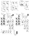
Figure 6. Tfh cells develop into effector Th2 cells after HDM challenge
(A-C) CD45.1+ B6 mice were irradiated and reconstituted with BM from Il21-mCherry and Il2-emGFP dual-reporter mice. Eight weeks later, reconstituted-chimeric mice were i.n HDM sensitized on day 1 and analyzed on day 6. The frequency of IL-21-expressing (mCherry+) and IL-2-expressing (GFP+) cells in CD44hiCD4+ T cells from the mLN (A) and lung (C) and are shown. Expression of intracellular IL-21, CXCR5 and PD1 in mCherry+ GFP single-positive and mCherry GFP+ single-positive CD4+ T cells from the mLN (B). (D-N) 1×10⁵ mCherry+ GFP single-positive (CXCR5+PD1+) or mCherry GFP+ single-positive (CXCR5PD1) CD44hiCD4+ T cells (D) or naïve CD44loCD4+ T cells, sorted by flow cytometry on day 6 after sensitization, were adoptively transferred into day 6 HDM sensitized CD45.1+ B6 mice. Recipient mice were then challenged with HDM or PBS on day 15 and analyzed on day 20. The frequency (E and I) and number (F and J) of donor CD45.2+ T cells in the mLN (E-F) and lung (I-J) are shown. The frequency of CXCR5 and PD1 (G and K) and mCherry-IL-21, GFP-IL-2 and intracellular IL-21 (H and L) in donor CXCR5hiPD1hi (Tfh) and CXCR5loPD1lo (Teff) CD4+ T cells from mLN (G-H) and lung (K-L) are shown. (M-N) The frequency (M) and number (N) of IL-4 and IL-13 double-producing cells among the donor CD45.2+ T cells were determined in the lungs by intracellular staining after restimulation for 4 h with anti-CD3 and BFA. (mean and S.D. of 4-5 mice per group).
Figure 7
Figure 7. Blockade of Tfh cell development impairs effector Th2 cell responses to HDM
(A-F) B6.4get mice were i.n HDM sensitized on day 1 and daily i.p treated with 50 mg/Kg of BCL-6 inhibitor 79-6 or vehicle (10% DMSO) from day 4 to day 11. Number of IL-4-expressing (EGFP+) Tfh cells from the mLN on day 12 (B). CD4+ T cells were purified from the mLN on day 12 and 2 × 10⁶ of purified CD4+ T cells were transferred into day 12 HDM-sensitized B6.CD45.1+ recipient mice. Recipient mice were i.n challenged on day 13 and analyzed on day 18 (A). Frequency (C) and number (D) of donor IL-4-expressing (EGFP+) CD4+ T cells in the lung. Frequency (E) and number (F) of donor IL-13+ CD4+ T cells in the lung. (G-O) 25×103 naïve OTII.CD45.1+ CD4+T cells were transferred into naïve congenic B6 mice on day 0. Mice were i.n HDM+OVA sensitized on day 1 and challenged on day 15. Mice were treated with 30.000 U of rIL-2 or PBS given twice a day for two days starting on day 10 or on day 16 (G). Number of donor Tfh OTII cells (H) and GC cells (I) in the mLN on day 12 Frequency (J) and number (K) of donor OTII cells from the lungs on day 20. Frequency (L) and number (M) of IL-4-producing cells among the donor OTII T cells determined in the lungs by intracellular staining after restimulation for 4 h with anti-CD3 and BFA. Number of eosinophils from day 20 lungs (N) and BAL (O). *P < 0.005 vs Ctrl. (unpaired Student’s t test). Data are representative of two independent experiments (mean and S.D. of 4-5 mice per group) See also Figure S4.

Comment in

References

    1. Ballesteros-Tato A, Leon B, Graf BA, Moquin A, Adams PS, Lund FE, Randall TD. Interleukin-2 inhibits germinal center formation by limiting T follicular helper cell differentiation. Immunity. 2012;36:847–856. - PMC - PubMed
    1. Ballesteros-Tato A, Randall TD. Priming of T follicular helper cells by dendritic cells. Immunology and cell biology. 2014;92:22–27. - PMC - PubMed
    1. Baumjohann D, Preite S, Reboldi A, Ronchi F, Ansel KM, Lanzavecchia A, Sallusto F. Persistent antigen and germinal center B cells sustain T follicular helper cell responses and phenotype. Immunity. 2013;38:596–605. - PubMed
    1. Cerchietti LC, Ghetu AF, Zhu X, Da Silva GF, Zhong S, Matthews M, Bunting KL, Polo JM, Fares C, Arrowsmith CH, et al. A small-molecule inhibitor of BCL6 kills DLBCL cells in vitro and in vivo. Cancer cell. 2010;17:400–411. - PMC - PubMed
    1. Choi YS, Kageyama R, Eto D, Escobar TC, Johnston RJ, Monticelli L, Lao C, Crotty S. ICOS receptor instructs T follicular helper cell versus effector cell differentiation via induction of the transcriptional repressor Bcl6. Immunity. 2011;34:932–946. - PMC - PubMed

Publication types

MeSH terms