. 2016 Mar 31;127(13):1666-75.
doi: 10.1182/blood-2015-12-684399. Epub 2016 Jan 29.

Janus kinase inhibition lessens inflammation and ameliorates disease in murine models of hemophagocytic lymphohistiocytosis

Affiliations

Janus kinase inhibition lessens inflammation and ameliorates disease in murine models of hemophagocytic lymphohistiocytosis

Rupali Das et al. Blood. .

Abstract

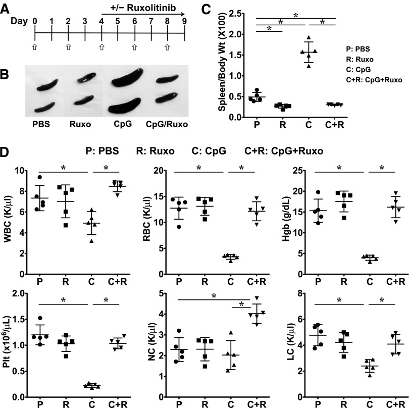
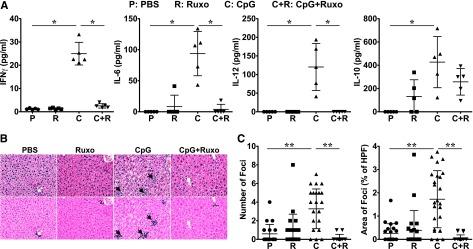
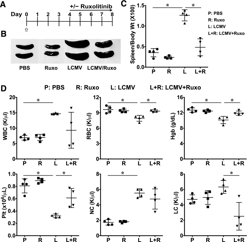
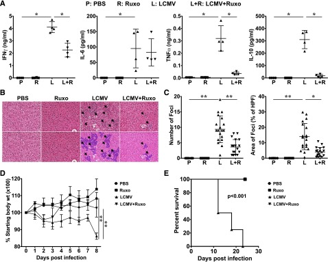
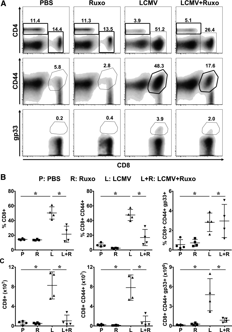
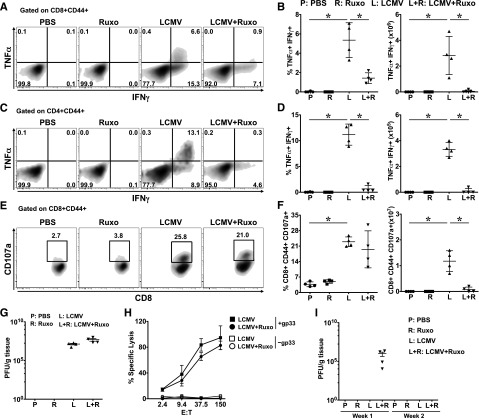
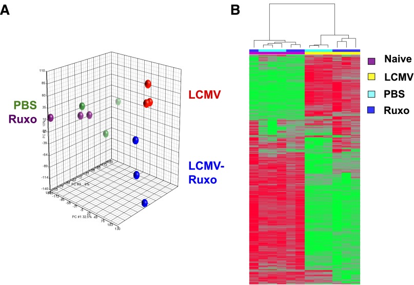
Hemophagocytic lymphohistiocytosis (HLH) comprises an emerging spectrum of inherited and noninherited disorders of the immune system characterized by the excessive production of cytokines, including interferon-γ and interleukins 2, 6, and 10 (IL-2, IL-6, and IL-10). The Janus kinases (JAKs) transduce signals initiated following engagement of specific receptors that bind a broad array of cytokines, including those overproduced in HLH. Based on the central role for cytokines in the pathogenesis of HLH, we sought to examine whether the inhibition of JAK function might lessen inflammation in murine models of the disease. Toward this end, we examined the effects of JAK inhibition using a model of primary (inherited) HLH in which perforin-deficient (Prf1(-∕-)) mice are infected with lymphocytic choriomeningitis virus (LCMV) and secondary (noninherited) HLH in which C57BL/6 mice receive repeated injections of CpG DNA. In both models, treatment with the JAK1/2 inhibitor ruxolitinib significantly lessened the clinical and laboratory manifestations of HLH, including weight loss, organomegaly, anemia, thrombocytopenia, hypercytokinemia, and tissue inflammation. Importantly, ruxolitinib treatment also significantly improved the survival of LCMV-infectedPrf1(-∕-)mice. Mechanistic studies revealed that in vivo exposure to ruxolitinib inhibited signal transducer and activation of transcription 1-dependent gene expression, limited CD8(+)T-cell expansion, and greatly reduced proinflammatory cytokine production, without effecting degranulation and cytotoxic function. Collectively, these findings highlight the JAKs as novel, druggable targets for mitigating the cytokine-driven hyperinflammation that occurs in HLH. These observations also support the incorporation of JAK inhibitors such as ruxolitinib into future clinical trials for patients with these life-threatening disorders.


Figures

Figure 1
Figure 1
Treatment with ruxolitinib lessens CpG-induced splenomegaly and cytopenias. (A) C57BL/6 (B6) mice were treated with PBS or CpG (50 μg) every other day as indicated (open/white arrow). Beginning on day 4, mice did or did not receive ruxolitinib (Ruxo) twice daily by oral gavage. On day 9, mice were euthanized and analyses performed. (B) Whole-spleen images from treatment groups. (C) Splenomegaly was quantified by measuring the ratio of spleen to body weight × 100. (D) Blood was analyzed for WBCs, RBCs, hemoglobin, platelets, neutrophils (NC), and lymphocytes (LC). Individual symbols each depict 1 mouse with horizontal lines representing the mean ± standard deviation (SD). Data are representative of 3 independent experiments. *P < .05.
Figure 2
Figure 2
Ruxolitinib treatment reduces CpG-induced hypercytokinemias and ameliorates liver inflammation. (A) Serum cytokine levels were assessed on day 9. (B) H&E-stained liver sections demonstrate inflammatory infiltrates (dark purple clusters), indicated by arrows. Representative sections are shown at a magnification of ×20 (top panels) and following computer analysis of inflammatory area (bottom panels). (C) The number of inflammatory foci (left) and percent area occupied by inflammatory foci with respect to the high-power field (HPF) of view (right) was determined by computer analysis of histologic samples. Symbols in panel A represent individual mice in each treatment group where in panel C they represent the number of, or area encompassed by, inflammatory foci (clusters containing >8 lymphocytes) per ×20 field of view. Data shown are mean ± SD and are representative of 3 independent experiments. *P < .05; **P < .001.
Figure 3
Figure 3
Treatment with ruxolitinib improves laboratory features of HLH in LCMV-infected Prf1−∕− mice. (A) Prf1−∕− mice were infected with 2 × 10⁵ PFU LCMV on day 0 (open/white arrow). Starting on day 4, mice were treated or not with ruxolitinib twice daily by oral gavage. Between days 8 and 10, mice were euthanized and analyses performed. (B) Whole-spleen images from treatment groups. (C) Splenomegaly was quantified by measuring the ratio of spleen to body weight × 100. (D) Blood was analyzed for the number of WBCs, RBCs, Hgb, Plt, NCs, and LCs. Data shown are mean ± SD and are representative of 4 independent experiments. *P < .05.
Figure 4
Figure 4
Ruxolitinib treatment reduces LCMV-induced hypercytokinemias and ameliorates liver inflammation. (A) Serum cytokine levels were assessed on days 8 to 10. (B) H&E-stained liver sections demonstrate inflammatory infiltrates (dark purple clusters), indicated by arrows. Representative sections are shown at a magnification of ×20 (top panels) and following computer analysis of inflammatory area (bottom panels). (C) The number of inflammatory foci (left) and percent area occupied by inflammatory foci with respect to the HPF view (right) was determined as in Figure 2. Data shown as mean ± SD are representative of 4 experiments. (D) Daily body weight is depicted as a ratio of the current over initial body weight ×100 with statistical significance determined by 2-way ANOVA. Symbols in panel A represent individual mice in each treatment group; in panel C, they represent the number of, or area encompassed by, inflammatory foci (clusters containing >8 lymphocytes) per ×20 field of view, whereas in panel D they represent the mean daily weight of the animals in each cohort. (E) Overall survival is depicted and statistical significance was determined by the log-rank test. Data in panels D and E are representative of 4 and 2 independent experiments, respectively. *P < .05, **P < .001.
Figure 5
Figure 5
Ruxolitinib reduces the numbers of activated and LCMV-specific CD8+ T cells in LCMV-infected Prf1−∕− mice. Mice treated with PBS, ruxolitinib, LCMV, and LCMV + ruxolitinib (L+R) were euthanized on day 9 postinfection. Splenocytes were stained using anti-CD4, CD8, CD44 antibodies and fluorochrome-conjugated MHC class I tetramer (Dbgp33). (A) Representative flow cytometric plots showing the percentages of CD4+, CD8+, CD8+CD44+ (top and middle panels, gated on live splenocytes) and CD8+gp33+ (bottom panels, gated on CD8+CD44+ cells) cells from a representative mouse in each group. (B) Percentage and (C) absolute number of splenic CD8+, CD8+CD44+, and CD8+CD44+gp33+ cells. Data are shown as mean ± SD and are representative of 4 independent experiments. *P < .05.
Figure 6
Figure 6
In vivo exposure to ruxolitinib differentially influences T-cell functions. Prf1−∕− mice were infected with LCMV and treated with ruxolitinib as described. On day 9, splenocytes from these animals were restimulated ex vivo with MHC class I (gp33-41)- or class II (gp61-80)-restricted LCMV peptides. Percentages of CD8+CD44+ (A-B [left]) and CD4+CD44+ (C-D [left]) T cells producing both TNFα and IFNγ, or CD8+CD44+ T cells expressing LAMP1 (CD107a; E-F [left]) were determined by intracellular and surface staining and flow cytometry. Absolute number of CD8+CD44+ and CD4+CD44+ T cells producing both TNFα and IFNγ, and CD8+CD44+CD107+ are shown in panels B, D, and F, respectively (right panels). In panels B, D, and F, data are presented as mean ± SD. (G) Viral titer was determined in the liver samples of Prf1−∕− mice in each group. (H) EL4 cells were pulsed with 20 μM gp33-41 peptide or left untreated and used as targets in an in vitro cytotoxicity assay. Cytolysis of gp33-loaded (filled symbols) or unloaded (open symbols) EL4 cells by splenic CD8+CD44+ cells of LCMV-infected or LCMV + ruxolitinib–treated (L+R) B6 mice. Specific lysis was determined by 51Cr release and plotted as the mean ± SD. Statistical significance in percent-specific lysis was determined by 2-way ANOVA. (I) Viral titers in the livers of B6 mice on day 8 (week 1) and day 15 (week 2) postinfection. Data in panels A through G and H and I are representative of 4 and 2 independent experiments, respectively. *P < .05.
Figure 7
Figure 7
Ruxolitinib treatment inhibits STAT1-dependent gene expression in splenic CD8+ T cells from LCMV-infected Prf1−/− mice. TCRβ+ CD8+CD44+ T cells were sort-purified from the spleens of uninfected untreated (PBS), uninfected ruxolitinib-treated (Ruxo), LCMV-infected (LCMV), and LCMV-infected ruxolitinib-treated (LCMV, Ruxo) Prf1−∕− mice. Sorted T cells were used to isolate RNA and complete complementary DNA microarray analysis. (A) PCA of the unfiltered data set. Each dot represents 1 mouse within each cohort. (B) Unsupervised hierarchical clustering of probe sets filtered on a log2 expression difference of >1.5 (1035 total probe sets). The height of the branches in the dendrogram reflects the distances between samples. Probe sets in green represent upregulated genes, compared with the mean expression; red represents downregulated genes.

Comment in

References

    1. Janka GE. Familial and acquired hemophagocytic lymphohistiocytosis. Annu Rev Med. 2012;63:233–246. - PubMed
    1. Canna SW, Behrens EM. Making sense of the cytokine storm: a conceptual framework for understanding, diagnosing, and treating hemophagocytic syndromes. Pediatr Clin North Am. 2012;59(2):329–344. - PMC - PubMed
    1. Janka GE, Lehmberg K. Hemophagocytic syndromes--an update. Blood Rev. 2014;28(4):135–142. - PubMed
    1. Trottestam H, Horne A, Aricò M, et al. Histiocyte Society. Chemoimmunotherapy for hemophagocytic lymphohistiocytosis: long-term results of the HLH-94 treatment protocol. Blood. 2011;118(17):4577–4584. - PMC - PubMed
    1. Marsh RA, Allen CE, McClain KL, et al. Salvage therapy of refractory hemophagocytic lymphohistiocytosis with alemtuzumab. Pediatr Blood Cancer. 2013;60(1):101–109. - PMC - PubMed

Publication types

MeSH terms