Review
. 2016 Mar;21(1):13-28.
doi: 10.1007/s00775-016-1336-0. Epub 2016 Jan 29.

Ferritins: furnishing proteins with iron

Affiliations
Review

Ferritins: furnishing proteins with iron

Justin M Bradley et al. J Biol Inorg Chem. 2016 Mar.

Abstract

Ferritins are a superfamily of iron oxidation, storage and mineralization proteins found throughout the animal, plant, and microbial kingdoms. The majority of ferritins consist of 24 subunits that individually fold into 4-α-helix bundles and assemble in a highly symmetric manner to form an approximately spherical protein coat around a central cavity into which an iron-containing mineral can be formed. Channels through the coat at inter-subunit contact points facilitate passage of iron ions to and from the central cavity, and intrasubunit catalytic sites, called ferroxidase centers, drive Fe(2+) oxidation and O2 reduction. Though the different members of the superfamily share a common structure, there is often little amino acid sequence identity between them. Even where there is a high degree of sequence identity between two ferritins there can be major differences in how the proteins handle iron. In this review we describe some of the important structural features of ferritins and their mineralized iron cores, consider how iron might be released from ferritins, and examine in detail how three selected ferritins oxidise Fe(2+) to explore the mechanistic variations that exist amongst ferritins. We suggest that the mechanistic differences reflect differing evolutionary pressures on amino acid sequences, and that these differing pressures are a consequence of different primary functions for different ferritins.

Keywords: Bacterioferritin; Ferritin; Ferroxidase; Iron oxidation; Iron storage.


Figures

Fig. 1
Fig. 1
Overall structure of 24meric ferritins. Left, view down one of six fourfold channels through the protein coat. The locations of four of the 24 B-channels are indicated by ‘B’. Right, view down one of eight threefold channels. Generated using PyMol with PDB file 1BCF
Fig. 2
Fig. 2
Comparison of B-channels in wild-type E. coli BFR and its D132F variant. One of the B-type channels formed at the interface between three subunits is displayed with the separate subunits coloured magenta, cyan and green. The amino acids forming the B-channels and the molecular surfaces generated with a 0.8 Å solvent probe radius (to mimic the hydrated Fe2+ substrate) are displayed for wild-type BFR (a, c) and its D132F variant (b, d). The D132F variant was constructed along with other variants involving residues lining the B channels to investigate whether the B channels are important for iron core formation [16] Constructed from PDB 1D3E1L. Reproduced with permission from Wong et al. [16]
Fig. 3
Fig. 3
The ferroxidase center of EcBFR. a The diiron ferroxidase center of BFR is shown with coordinating residues (Glu18 and His54 are terminal ligands to Fe1; Glu94 and His130 are terminal ligands to Fe2; Glu51 and Glu127 bridge Fe1 and Fe2), along with the inner surface iron site (FeIS) with coordinating residues (His46 and Asp50), and closely lying aromatic residues (Tyr25, Tyr58 and Trp133). b The apo-form of the ferroxidase center of BFR showing that residues that act as ligands to the irons are located in similar positions, with the exception of His130, which, in this apo-structure, has swung away from the iron binding sites such that the center adopts an open conformation. a and b generated using PyMol with PDB files 3E1M and 3E1L, respectively
Fig. 4
Fig. 4
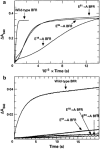
Kinetic traces for Fe oxidation in EcBFR. a Absorption change at 340 nm measured as a function of time after the addition of 400 Fe2+ ions per apo-protein molecule to samples of wild-type, E18A, E51A and E94A BFR, as indicated. For the wild-type protein this profile yields the phase 3 rate. b Absorption change at 340 nm followed by spectrophotometry over the first 20 s following the addition of 400 Fe2+ ions per apo-protein molecule. For the wild-type protein this profile yields the phase 2 rate. Proteins were in 100 mM MES buffer, pH 6.5 at a final concentration of 0.5 µM. Temperature was 30 °C, pathlength 1 cm. Reproduced with permission from Le Brun et al. [22]
Fig. 5
Fig. 5
Summary of the proposed BFR mineralization mechanism. Two Fe2+ ions access the ferroxidase site via B-type channels [16] and undergo oxidation to the bridged di-ferric form (with either O2 or H2O2 as oxidant [75]. The oxidized di-Fe3+ form of the site is stable [23]. Additional Fe2+ ions binds at the inner surface site (IS site) and undergoes oxidation to Fe3+. A second electron is derived from the oxidation of the nearby Tyr25 side chain, generating a radical and regenerating the di-Fe2+ form of the ferroxidase site. The radical decays indirectly through the oxidation of a second Fe2+ ion (at an unknown location), and the oxidized iron at the inner surface site nucleates or is incorporated into the growing mineral core. The di-Fe2+ ferroxidase site undergoes oxidation again via reaction with O2 or H2O2. At this point, the catalytic site has returned to its resting state, ready to react again when Fe2+ ions are present. Hydrolysis of the accumulating hydrated Fe3+ ions in the cavity leads to mineral formation. Adapted from Bradley et al. [76]
Fig. 6
Fig. 6
The Ftn-type ferroxidase center and site C of P. multiseries Ftn. Structure of the eukaryotic Ftn-type ferroxidase center and site C of P. multiseries Ftn with Fe3+ ions bound generated using PyMol from PDB 4IWK [79]
Fig. 7
Fig. 7
Stopped -flow spectroscopy of iron mineralization in wild type and variant PmFTN Absorbance changes at 340 nm showing Fe2+ oxidation following addition of 400 Fe2+/PmFTN to wild type and variant PmFTN (0.5 μM) in 0.1 MES pH 6.5 at 25 °C. The profile of the plots from ~50 s onwards is determined by the rate of the phase 3 reactions. With the E130A variant this is complete in about 300 s but with the wild-type protein it is only about 40 % done in 1000 s. Note that in EcBFR under similar conditions the phase 3 reaction with 400 Fe2+/EcBFR is complete in about 1000 s (Fig. 4a). Reproduced with permission from Pfaffen et al. [80]

References

    1. Laufberger V. Bull Soc Chim biol. 1937;19:1575–1582.
    1. Lewis CT, Short C (1879) A latin dictionary. Clarendon Press, Oxford. http://www.perseus.tufts.edu. Accessed 3 Jan 2016
    1. Ford GC, Harrison PM, Rice DW, Smith JMA, Treffry A, White JL, Yariv J. Philos Trans R Soc Lond Ser. 1984;B304:551–565. doi: 10.1098/rstb.1984.0046. - DOI - PubMed
    1. Harrison PM, Arosio P. Biochim Biophys Acta. 1996;1275:161–203. doi: 10.1016/0005-2728(96)00022-9. - DOI - PubMed
    1. Lewin A, Moore GR, Le Brun NE (2005) Dalton Trans, pp 3597–3610 - PubMed

Publication types

LinkOut - more resources