

Effect of biosurfactants on Pseudomonas aeruginosa and Staphylococcus aureus biofilms in a BioFlux channel
- PMID: 26825819
- PMCID: PMC4909806
- DOI: 10.1007/s00253-016-7310-5
Effect of biosurfactants on Pseudomonas aeruginosa and Staphylococcus aureus biofilms in a BioFlux channel
Abstract
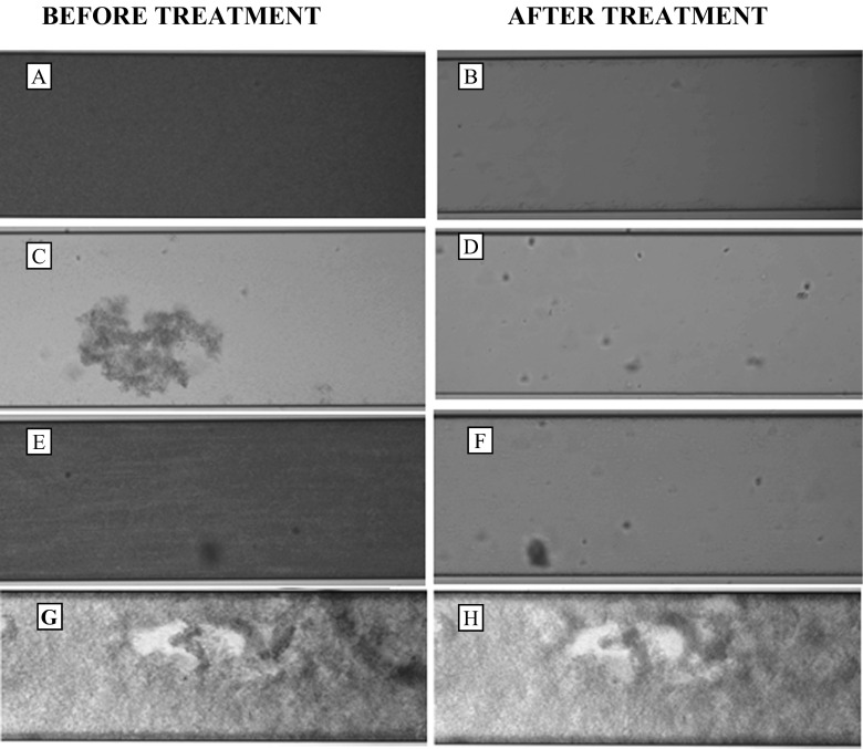
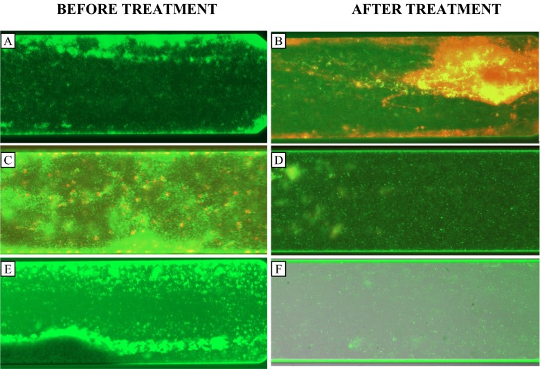
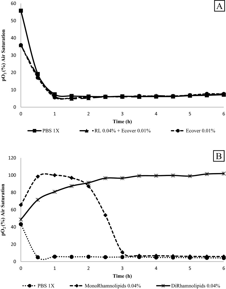
Recent studies have indicated that biosurfactants play a role both in maintaining channels between multicellular structures in biofilms and in dispersal of cells from biofilms. A combination of caprylic acid (0.01 % v/v) together with rhamnolipids (0.04 % v/v) was applied to biofilms of Pseudomonas aeruginosa ATCC 15442, Staphylococcus aureus ATCC 9144 and a mixed culture under BioFlux flowthrough conditions and caused disruption of the biofilms. The biofilms were also treated with a combination of rhamnolipids (0.04 % v/v) and sophorolipids (0.01 %). Control treatments with PBS 1× had no apparent effect on biofilm disruption. The Gram-positive bacterium (S. aureus ATCC 9144) was more sensitive than P. aeruginosa ATCC 15442 in terms of disruption and viability as shown by Live/Dead staining. Disruption of biofilms of P. aeruginosa ATCC 15442 was minimal. Oxygen consumption by biofilms, after different treatments with biosurfactants, confirms that sophorolipid on its own is unable to kill/inhibit cells of P. aeruginosa ATCC 15442, and even when used in combination with rhamnolipids, under static conditions, no decrease in the cell viability was observed. Cells in biofilms exposed to mono-rhamnolipids (0.04 % v/v) showed behaviour typical of exposure to bacteriostatic compounds, but when exposed to di-rhamnolipids (0.04 % v/v), they displayed a pattern characteristic of bactericidal compounds.
Keywords: BioFlux; Biofilms; Biosurfactants; Rhamnolipids.
Figures
Similar articles
-
Pseudomonas aeruginosa biofilm disruption using microbial surfactants.J Appl Microbiol. 2016 Apr;120(4):868-76. doi: 10.1111/jam.13049. Epub 2016 Feb 18. J Appl Microbiol. 2016. PMID: 26742560
-
Antibacterial properties of biosurfactants against selected Gram-positive and -negative bacteria.FEMS Microbiol Lett. 2016 Jan;363(2):fnv224. doi: 10.1093/femsle/fnv224. Epub 2015 Nov 22. FEMS Microbiol Lett. 2016. PMID: 26598715
-
Effect of Mono and Di-rhamnolipids on Biofilms Pre-formed by Bacillus subtilis BBK006.Curr Microbiol. 2016 Aug;73(2):183-9. doi: 10.1007/s00284-016-1046-4. Epub 2016 Apr 25. Curr Microbiol. 2016. PMID: 27113589 Free PMC article.
-
The involvement of rhamnolipids in microbial cell adhesion and biofilm development - an approach for control?Lett Appl Microbiol. 2014 May;58(5):447-53. doi: 10.1111/lam.12211. Epub 2014 Jan 24. Lett Appl Microbiol. 2014. PMID: 24372465 Review.
-
[Advances in the study of synergistic effect of anti-biofilm agents].Yao Xue Xue Bao. 2012 Mar;47(3):339-45. Yao Xue Xue Bao. 2012. PMID: 22645757 Review. Chinese.
Cited by
-
Natural Medicine a Promising Candidate in Combating Microbial Biofilm.Antibiotics (Basel). 2023 Feb 2;12(2):299. doi: 10.3390/antibiotics12020299. Antibiotics (Basel). 2023. PMID: 36830210 Free PMC article. Review.
-
Harnessing the Potential of Biosurfactants for Biomedical and Pharmaceutical Applications.Pharmaceutics. 2023 Aug 18;15(8):2156. doi: 10.3390/pharmaceutics15082156. Pharmaceutics. 2023. PMID: 37631370 Free PMC article. Review.
-
Sophorolipid: An Effective Biomolecule for Targeting Microbial Biofilms.Curr Microbiol. 2024 Oct 4;81(11):388. doi: 10.1007/s00284-024-03892-6. Curr Microbiol. 2024. PMID: 39367190 Review.
-
Towards individualized diagnostics of biofilm-associated infections: a case study.NPJ Biofilms Microbiomes. 2017 Sep 28;3:22. doi: 10.1038/s41522-017-0030-5. eCollection 2017. NPJ Biofilms Microbiomes. 2017. PMID: 28970943 Free PMC article.
-
Ultrasonic preparation, structural characterization and functional enhancement of thyme essential oil-loaded microcapsules based on mannosylerythritol lipid-A.Ultrason Sonochem. 2025 Mar;114:107265. doi: 10.1016/j.ultsonch.2025.107265. Epub 2025 Feb 9. Ultrason Sonochem. 2025. PMID: 39933309 Free PMC article.
References
-
- Callaghan B, Roelants S, Baccile N, Lydon H, Van Bogaert I, Banat IM, Marchant R, Mitchell CA (2015) Sophorolipid-mediated inhibition of colorectal tumor cell growth in vitro and in vivo Cancer Res 75(15):Abstract nr 2294. doi:10.1158/1538–7445.AM2015-2294
-
- Díaz De Rienzo MA, Stevenson P, Marchant R, Banat IM. Antibacterial properties of biosurfactants against selected gram positive and negative bacteria. FEMS Microbiol Lett. 2015 - PubMed
MeSH terms
Substances
LinkOut - more resources
Full Text Sources
Other Literature Sources
Medical
Molecular Biology Databases
