. 2017 Feb 1;27(2):1670-1685.
doi: 10.1093/cercor/bhw009.

Cc2d1a Loss of Function Disrupts Functional and Morphological Development in Forebrain Neurons Leading to Cognitive and Social Deficits

Affiliations

Cc2d1a Loss of Function Disrupts Functional and Morphological Development in Forebrain Neurons Leading to Cognitive and Social Deficits

Adam W Oaks et al. Cereb Cortex. .

Abstract

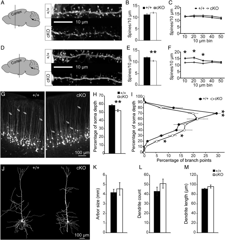
Loss-of-function (LOF) mutations in CC2D1A cause a spectrum of neurodevelopmental disorders, including intellectual disability, autism spectrum disorder, and seizures, identifying a critical role for this gene in cognitive and social development. CC2D1A regulates intracellular signaling processes that are critical for neuronal function, but previous attempts to model the human LOF phenotypes have been prevented by perinatal lethality in Cc2d1a-deficient mice. To overcome this challenge, we generated a floxed Cc2d1a allele for conditional removal of Cc2d1a in the brain using Cre recombinase. While removal of Cc2d1a in neuronal progenitors using Cre expressed from the Nestin promoter still causes death at birth, conditional postnatal removal of Cc2d1a in the forebrain via calcium/calmodulin-dependent protein kinase II-alpha (CamKIIa) promoter-driven Cre generates animals that are viable and fertile with grossly normal anatomy. Analysis of neuronal morphology identified abnormal cortical dendrite organization and a reduction in dendritic spine density. These animals display deficits in neuronal plasticity and in spatial learning and memory that are accompanied by reduced sociability, hyperactivity, anxiety, and excessive grooming. Cc2d1a conditional knockout mice therefore recapitulate features of both cognitive and social impairment caused by human CC2D1A mutation, and represent a model that could provide much needed insights into the developmental mechanisms underlying nonsyndromic neurodevelopmental disorders.

Keywords: autism; cognitive development; dendritic spines; intellectual disability; long-term plasticity.


Figures

Figure 1.
Figure 1.
Cc2d1a KO mice exhibit early postnatal lethality. (A) Gene trap containing En2SA sequence, and a separate neomycin resistance cassette is flanked by FRT sites and produces a transcript truncated between exons 11 and 12. LoxP sites for Cre recombinase targeting flank exons 12–14. (B) Domain organization of full length human and murine proteins (4 DM14 domains and 1 C2 domain) compared with a predicted protein product from a disease-causing CC2D1A mutation (Basel-Vanagaite et al. 2006) and the truncated proteins produced in our gene-trap mouse and a previously reported Cc2d1a null mouse (Al-Tawashi et al. 2012). (C) Immunoblot analysis of Cc2d1a expression in WT (+/+), heterozygous (+/−), and null mice (−/−) shows the complete loss of full length (fl) Cc2d1a protein. (D) Brains dissected from pups at embryonic day (E) 18.5 show now difference in size and organization is preserved in E18.5 brain tissue sections stained with H&E (E). (F) The weight of pups following Cesarean delivery at E18.5 is the same across genotypes. Results expressed as mean ± SEM of indicated number of pups from 4 litters. (G) Cc2d1a null mice are born in reduced numbers relative to predicted Mendelian ratios and do not survive beyond postnatal day (P) 0 (data from 5 to 10 litters per age).
Figure 2.
Figure 2.
Cc2d1a KO mice show breathing and swallowing deficits. (A) Ratio of pups breathing at birth following Cesarean delivery. (B) Ratio of pups that could be induced to swallow at birth following Cesarean delivery. Results expressed as the percent of total of pups from 4 litters (A and B). (C) Cc2d1a KO pups show accumulation of fluorescently labeled milk (in green; right panels) in the lungs (L) and stomach (S), while milk can only be found in the stomach in WTs.
Figure 3.
Figure 3.
Postnatal forebrain removal of Cc2d1a rescues lethality. (A) Removal of gene-trap allele by FRT following cross of Cc2d1a heterozygous mice with the FLPeR mouse line, leaving LoxP sites flanking exons 12–14 in Cc2d1aflx mice. (B) Cre recombinase expression under the CamKIIa promoter rescues early postnatal lethality (data from 16 litters), while crossing with Nestincre does not (data from 6 litters). (C) Cc2d1a is almost completely absent from the cortex of adult cKO mice, even though CamKIIcre is only expressed in excitatory neurons. (D) Analysis of steady-state Cc2d1a mRNA expression in Cc2d1aflx/flx (+/+) and cKO brain tissue also shows almost complete ablation of mRNA both upstream (5′) and downstream (3′) of the predicted truncation. Levels in cKO cortex (ctx) and hippocampus (hip) are expressed as fold change relative to respective WT tissues. Results expressed as mean from 4 mice per genotype, *P < 0.05, ***P < 0.001 (two-tailed t-tests in D). (E) The size and organization of the adult cKO brain is indistinguishable from WT littermates Cc2d1aflx/flx stained with H&E.
Figure 4.
Figure 4.
Dendrite and spine morphology is altered in the cortex of cKO mice. (A) No changes in dendritic spine (B) density and (C) distribution are observed on secondary apical dendrites on layer V cells of the anterior somatosensory cortex (Bregma −0.23 mm) in WT (+/+) and Cc2d1a cKO mice. (D) Dendritic spine density is reduced (E), especially in the initial segment of secondary dendrites (F) in more posterior somatosensory cortex (Bregma −1.55 mm). Results expressed as mean ± SEM of n = 5 mice per genotype (7–10 dendrites per animal), *P < 0.05, **P < 0.01 (two-tailed t-tests in C and F). (G) The branching point of the apical tuft in layer V neurons of the posterior somatosensory cortex (bifurcations marked by arrows) was found to be lower in cKO brains, as indicated by (H) population average and (I) distribution of branch depths relative to soma depth (0% = closest to soma; 100% = furthest from soma). Results expressed as mean ± SEM of n = 5 mice per genotype (100–200 branch points per animal), *P < 0.05, **P < 0.01 (two-tailed t-tests in H and I). (J) No difference was observed in dendrite (K) size, (L) number, and (M) average length in the dendritic arbor of Golgi-stained layer V neurons of the posterior somatosensory cortex in WT (+/+) and Cc2d1a cKO mice. Results expressed as mean ± SEM of n = 4 mice per genotype (6–8 arbor traces per animal; two-tailed t-tests in K, L, and M).
Figure 5.
Figure 5.
Hippocampus-dependent memory formation is impaired in the Cc2d1a cKO mice. (A) Short-term memory was examined by the NORT test, with a novel object replacing a familiar object after a 15-min intersession interval. In contrast to WT (+/+; n = 11), Cc2d1a cKO mice (n = 15) showed no preference for the novel object relative to a familiar object. Results expressed as mean ± SEM, *P < 0.05 (two-tailed t-tests in A). (B) Spatial learning was measured as latency to escape in 3 stages of the MWM with a visible platform (V; 2 days), hidden platform (H; 5 days), or the reversal (R; 2 days) of the hidden platform location. Cc2d1a cKO (n = 15) mice were significantly impaired in learning the location of the hidden platforms compared with WT mice (+/+; n = 14) over 4 trials per day per stage. Results expressed as mean ± SEM of mice from each genotype, *P < 0.05 (Mann–Whitney U-tests or two-tailed t-tests for stages with unequal or equal variance, respectively, in B, following detection of significant interaction by two-way ANOVA). (C and D) Dendritic spine density on secondary apical dendrites of CA1 pyramidal neurons was slightly reduced. Results expressed as mean ± SEM of n = 5 mice per genotype (8–10 dendrites per animal), ***P < 0.001 (effect of genotype by two-way ANOVA in D). (E and F) fEPSP recordings show a defect in L-LTP (F; 100 Hz for 1 s with 20 s interval; 10 WT and 11 Cc2c1a cKO hippocampal preparations), but not in E-LTP (E; 100 Hz for 1 s; 8 hippocampal preparations per genotype). Representative traces recorded (1) before and (2) after stimulus protocols are displayed (insets). (G) Averaging of the last 10 min of fEPSPs recorded in the L-LTP indicates a significant reduction in synaptic strength. Results expressed as mean ± SEM of 10 WT and 11 Cc2c1a cKO hippocampal preparations, **P < 0.01 (two-tailed t-test in H).
Figure 6.
Figure 6.
Social interaction deficits in Cc2d1a cKO mice. (AC) Sociability is reduced for cKO mice in all analyses. Social interaction was assessed in the three-chamber test (see Supplementary Fig. 6A) and is presented as (A) time spent in each chamber, (B) entries to the perimeter around each cup, and (C) time spent interacting with each cup. Results expressed as mean ± SEM of 10 mice per genotype; *P < 0.05, **P < 0.01, ***P < 0.001 (two-tailed t-test within the genotype); #P < 0.05, ##P < 0.01, #P < 0.001 (two-tailed t-test between genotypes). Male–female social interaction was assessed during interaction between virgin WT (+/+) or cKO males and virgin WT females as (D) total social sniffing time which is the sum of (E) nose-to-body, (F) nose-to-genital, and (G) nose-to-nose sniffing. Values calculated as a percentage of the total testing duration (300 s) are expressed as mean ± SEM of 8 mice per genotype; **P < 0.01 (two-tailed t-tests in DG). (H) Representative sonograms of USVs recorded during interaction between virgin WT (+/+) or cKO males and virgin WT females. (I) Average number of vocalizations per pairing recorded across 5 pairings. Results expressed as mean ± SEM of 8 mice per genotype; *P < 0.05 (Mann–Whitney U-test in I).
Figure 7.
Figure 7.
cKO mice display behavioral phenotypes consistent with ID/ASD. (A–D) Exploratory activity in a novel environment was assessed on the open field test. Representative paths from (A) WT (+/+; n = 14) and (B) cKO mice (n = 15) mice show a substantial increase in locomotion in cKO animals. (C) Total path length is significantly increased, while (D) time spent in the center zone is reduced showing anxiety. Results expressed as mean ± SEM of mice from each genotype, **P < 0.01, ***P < 0.001 (two-tailed t-tests in C and D). (E) In the marble burying test, digging activity is significantly reduced in cKO mice, while other activities, such as exploring, sitting, and grooming (+/+, n = 12; cKO, n = 18), are unaffected. Results expressed as mean ± SEM of mice from each genotype, *P < 0.05 (two-tailed t-tests in E).

Similar articles

Cited by

References

    1. Abrahams BS, Arking DE, Campbell DB, Mefford HC, Morrow EM, Weiss LA, Menashe I, Wadkins T, Banerjee-Basu S, Packer A. 2013. SFARI Gene 2.0: a community-driven knowledgebase for the autism spectrum disorders (ASDs). Mol Autism. 4:36. - PMC - PubMed
    1. Al-Tawashi A, Jung S, Liu D, Su B, Qin J. 2012. Protein implicated in nonsyndromic mental retardation regulates protein kinase A (PKA) activity. J Biol Chem. 287:14644–14658. - PMC - PubMed
    1. Basel-Vanagaite L, Attia R, Yahav M, Ferland RJ, Anteki L, Walsh CA, Olender T, Straussberg R, Magal N, Taub E et al. . 2006. The CC2D1A, a member of a new gene family with C2 domains, is involved in autosomal recessive non-syndromic mental retardation. J Med Genet. 43:203–210. - PMC - PubMed
    1. Betancur C. 2011. Etiological heterogeneity in autism spectrum disorders: more than 100 genetic and genomic disorders and still counting. Brain Res. 1380:42–77. - PubMed
    1. Bevins R, Besheer J. 2006. Object recognition in rats and mice: a one-trial non-matching-to-sample learning task to study “recognition memory”. Nat Protoc. 1:1306–1311. - PubMed

Publication types