

An Antibody Biosensor Establishes the Activation of the M1 Muscarinic Acetylcholine Receptor during Learning and Memory
- PMID: 26826123
- PMCID: PMC4861454
- DOI: 10.1074/jbc.M115.681726
An Antibody Biosensor Establishes the Activation of the M1 Muscarinic Acetylcholine Receptor during Learning and Memory
Abstract
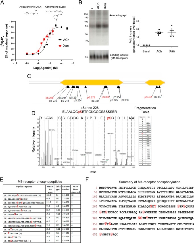
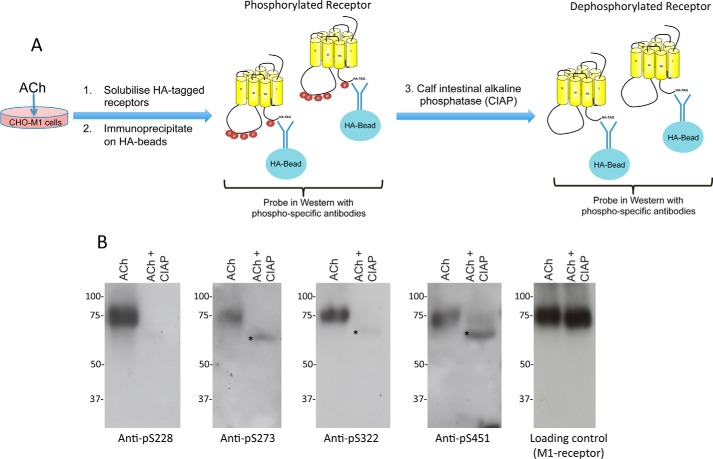
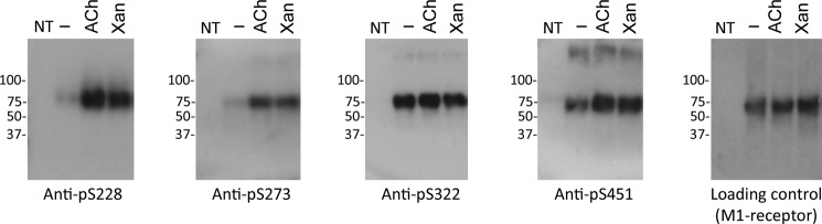
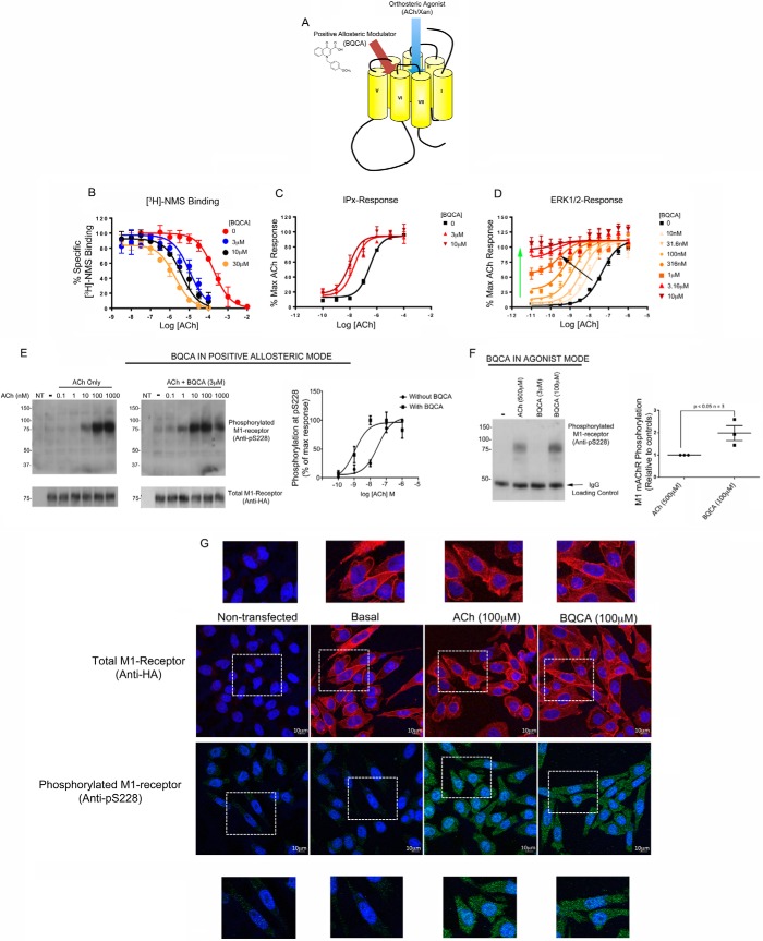
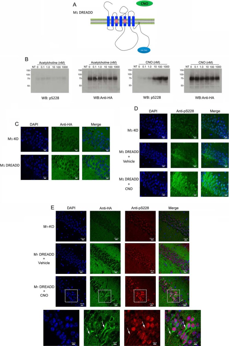
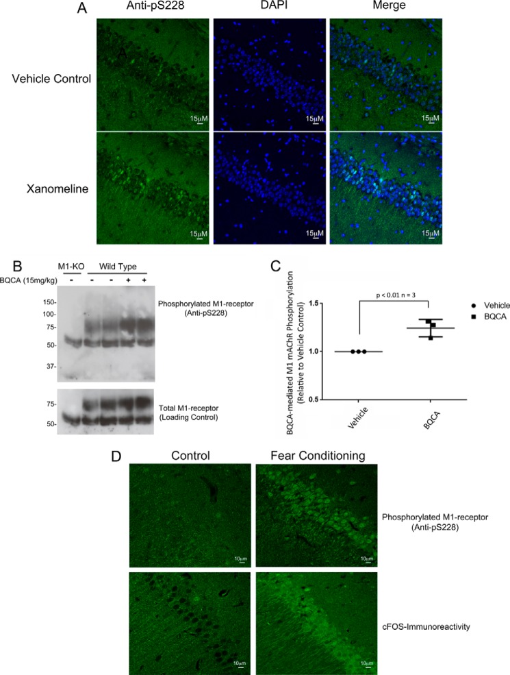
Establishing the in vivo activation status of G protein-coupled receptors would not only indicate physiological roles of G protein-coupled receptors but would also aid drug discovery by establishing drug/receptor engagement. Here, we develop a phospho-specific antibody-based biosensor to detect activation of the M1 muscarinic acetylcholine receptor (M1 mAChR) in vitro and in vivo Mass spectrometry phosphoproteomics identified 14 sites of phosphorylation on the M1 mAChR. Phospho-specific antibodies to four of these sites established that serine at position 228 (Ser(228)) on the M1 mAChR showed extremely low levels of basal phosphorylation that were significantly up-regulated by orthosteric agonist stimulation. In addition, the M1 mAChR-positive allosteric modulator, 1-(4-methoxybenzyl)-4-oxo-1,4-dihydroquinoline-3-carboxylic acid, enhanced acetylcholine-mediated phosphorylation at Ser(228) These data supported the hypothesis that phosphorylation at Ser(228) was an indicator of M1 mAChR activation. This was further supported in vivo by the identification of phosphorylated Ser(228) on the M1 mAChR in the hippocampus of mice following administration of the muscarinic ligands xanomeline and 1-(4-methoxybenzyl)-4-oxo-1,4-dihydroquinoline-3-carboxylic acid. Finally, Ser(228) phosphorylation was seen to increase in the CA1 region of the hippocampus following memory acquisition, a response that correlated closely with up-regulation of CA1 neuronal activity. Thus, determining the phosphorylation status of the M1 mAChR at Ser(228) not only provides a means of establishing receptor activation following drug treatment both in vitro and in vivo but also allows for the mapping of the activation status of the M1 mAChR in the hippocampus following memory acquisition thereby establishing a link between M1 mAChR activation and hippocampus-based memory and learning.
Keywords: G protein-coupled receptor (GPCR); drug discovery; hippocampus; learning; mass spectrometry (MS); memory; phosphorylation.
© 2016 by The American Society for Biochemistry and Molecular Biology, Inc.
Figures
Similar articles
-
Striatal, Hippocampal, and Cortical Networks Are Differentially Responsive to the M4- and M1-Muscarinic Acetylcholine Receptor Mediated Effects of Xanomeline.ACS Chem Neurosci. 2019 Mar 20;10(3):1753-1764. doi: 10.1021/acschemneuro.8b00625. Epub 2018 Dec 11. ACS Chem Neurosci. 2019. PMID: 30480428
-
Bitopic Binding Mode of an M1 Muscarinic Acetylcholine Receptor Agonist Associated with Adverse Clinical Trial Outcomes.Mol Pharmacol. 2018 Jun;93(6):645-656. doi: 10.1124/mol.118.111872. Epub 2018 Apr 25. Mol Pharmacol. 2018. PMID: 29695609 Free PMC article.
-
Molecular determinants of allosteric modulation at the M1 muscarinic acetylcholine receptor.J Biol Chem. 2014 Feb 28;289(9):6067-79. doi: 10.1074/jbc.M113.539080. Epub 2014 Jan 17. J Biol Chem. 2014. PMID: 24443568 Free PMC article.
-
M1 muscarinic acetylcholine receptors: A therapeutic strategy for symptomatic and disease-modifying effects in Alzheimer's disease?Adv Pharmacol. 2020;88:277-310. doi: 10.1016/bs.apha.2019.12.003. Epub 2020 Jan 27. Adv Pharmacol. 2020. PMID: 32416870 Review.
-
Development of M1 mAChR allosteric and bitopic ligands: prospective therapeutics for the treatment of cognitive deficits.ACS Chem Neurosci. 2013 Jul 17;4(7):1026-48. doi: 10.1021/cn400086m. Epub 2013 May 23. ACS Chem Neurosci. 2013. PMID: 23659787 Free PMC article. Review.
Cited by
-
Phosphorylation bar-coding of free fatty acid receptor 2 is generated in a tissue-specific manner.Elife. 2023 Dec 12;12:RP91861. doi: 10.7554/eLife.91861. Elife. 2023. PMID: 38085667 Free PMC article.
-
Selective phosphorylation of threonine residues defines GPR84-arrestin interactions of biased ligands.J Biol Chem. 2022 May;298(5):101932. doi: 10.1016/j.jbc.2022.101932. Epub 2022 Apr 12. J Biol Chem. 2022. PMID: 35427647 Free PMC article.
-
Muscarinic receptor regulates extracellular signal regulated kinase by two modes of arrestin binding.Proc Natl Acad Sci U S A. 2017 Jul 11;114(28):E5579-E5588. doi: 10.1073/pnas.1700331114. Epub 2017 Jun 26. Proc Natl Acad Sci U S A. 2017. PMID: 28652372 Free PMC article.
-
Grape Seed Proanthocyanidin and Swimming Exercise Protects Against Cognitive Decline: A Study on M1 Acetylcholine Receptors in Aging Male Rat Brain.Neurochem Res. 2017 Dec;42(12):3573-3586. doi: 10.1007/s11064-017-2406-6. Epub 2017 Oct 9. Neurochem Res. 2017. PMID: 28993969
-
Identification of Candidate Allosteric Modulators of the M1 Muscarinic Acetylcholine Receptor Which May Improve Vagus Nerve Stimulation in Chronic Tinnitus.Front Neurosci. 2017 Nov 14;11:636. doi: 10.3389/fnins.2017.00636. eCollection 2017. Front Neurosci. 2017. PMID: 29184482 Free PMC article.
References
-
- Jacoby E., Bouhelal R., Gerspacher M., and Seuwen K. (2006) The 7 TM G-protein-coupled receptor target family. ChemMedChem 1, 761–782 - PubMed
-
- Rasmussen S. G., DeVree B. T., Zou Y., Kruse A. C., Chung K. Y., Kobilka T. S., Thian F. S., Chae P. S., Pardon E., Calinski D., Mathiesen J. M., Shah S. T., Lyons J. A., Caffrey M., Gellman S. H., et al. (2011) Crystal structure of the β2 adrenergic receptor-Gs protein complex. Nature 477, 549–555 - PMC - PubMed
-
- Venkatakrishnan A. J., Deupi X., Lebon G., Tate C. G., Schertler G. F., and Babu M. M. (2013) Molecular signatures of G-protein-coupled receptors. Nature 494, 185–194 - PubMed
-
- Kenakin T. (2005) New concepts in drug discovery: collateral efficacy and permissive antagonism. Nat. Rev. Drug Discov. 4, 919–927 - PubMed
-
- Kenakin T., and Williams M. (2014) Defining and characterizing drug/compound function. Biochem. Pharmacol. 87, 40–63 - PubMed
Publication types
MeSH terms
Substances
Grants and funding
LinkOut - more resources
Full Text Sources
Other Literature Sources
Medical
Molecular Biology Databases
Miscellaneous
