. 2016 Mar 1;196(5):2401-9.
doi: 10.4049/jimmunol.1502470. Epub 2016 Jan 29.

Enhancement of Microbiota in Healthy Macaques Results in Beneficial Modulation of Mucosal and Systemic Immune Function

Affiliations

Enhancement of Microbiota in Healthy Macaques Results in Beneficial Modulation of Mucosal and Systemic Immune Function

Jennifer A Manuzak et al. J Immunol. .

Abstract

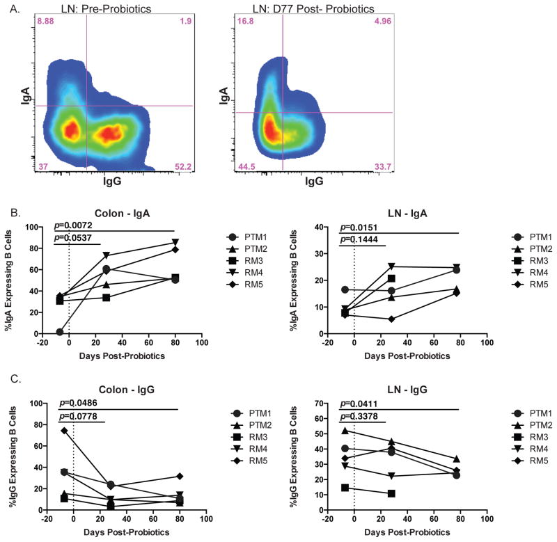
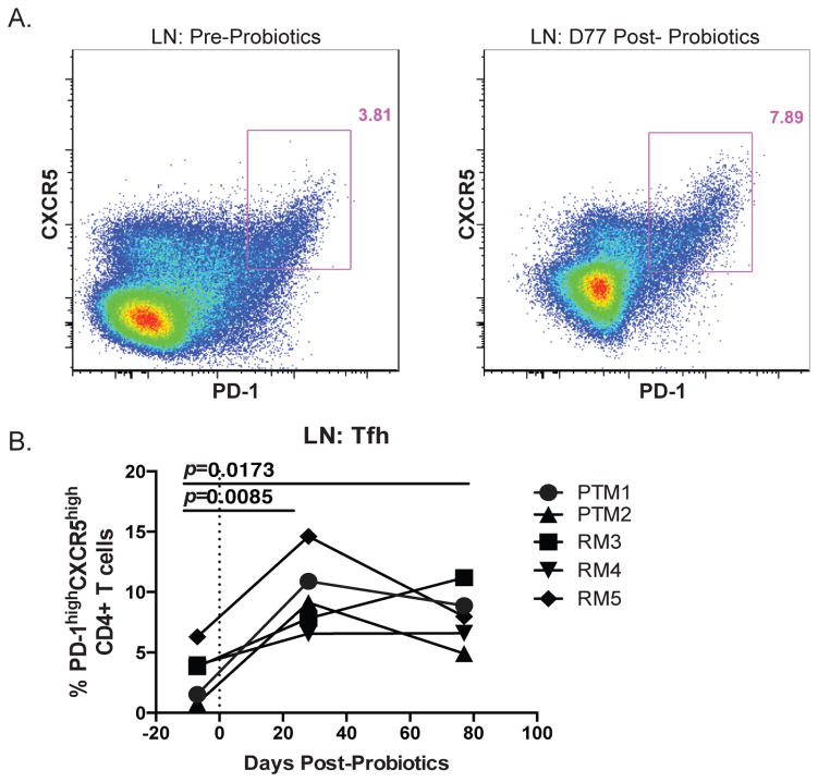
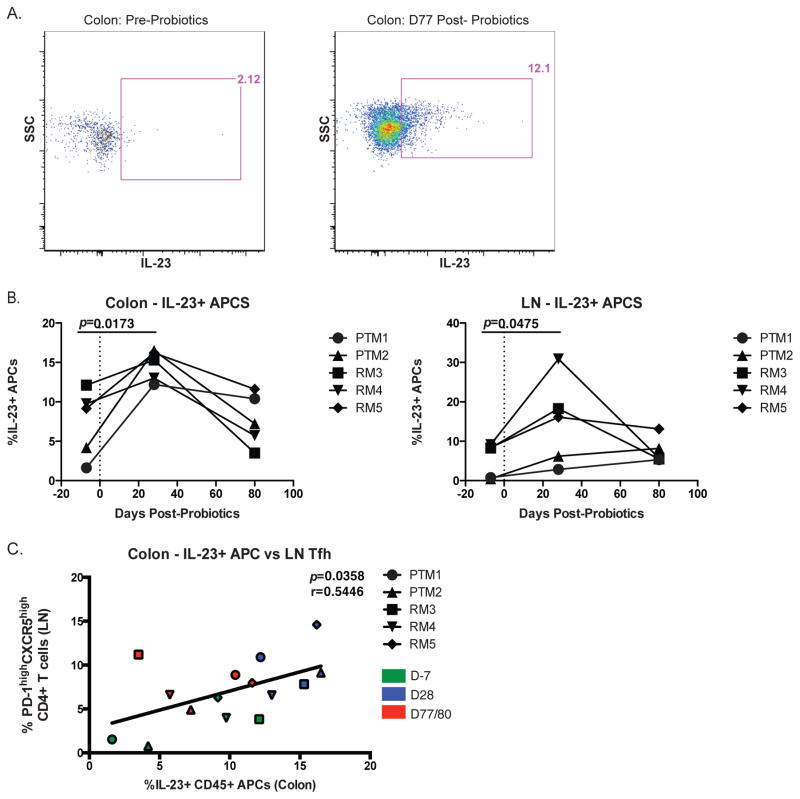
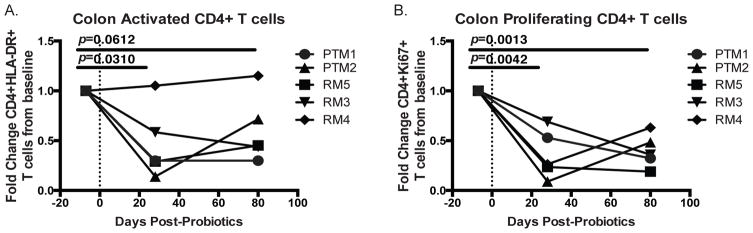
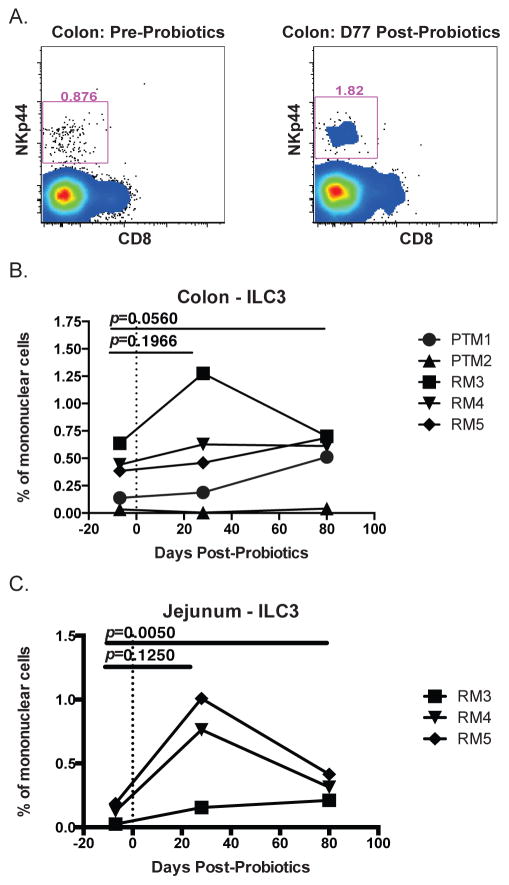
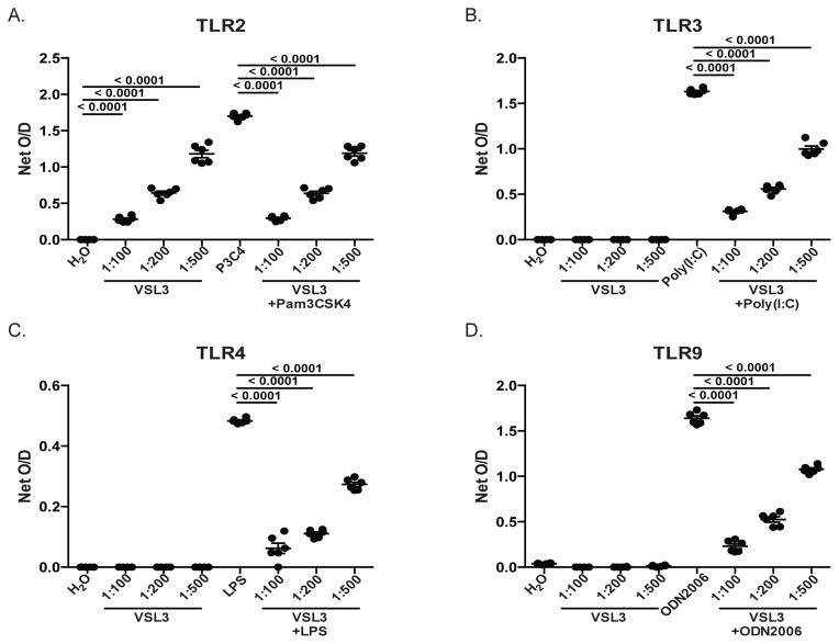
Given the critical role of mucosal surfaces in susceptibility to infection, it is imperative that effective mucosal responses are induced when developing efficacious vaccines and prevention strategies for infection. Modulating the microbiota in the gastrointestinal (GI) tract through the use of probiotics (PBio) is a safe and well-tolerated approach to enhance mucosal and overall health. We assessed the longitudinal impact of daily treatment with the VSL#3 probiotic on cellular and humoral immunity and inflammation in healthy macaques. PBio therapy resulted in significantly increased frequencies of B cells expressing IgA in the colon and lymph node (LN), likely because of significantly increased LN T follicular helper cell frequencies and LN follicles. Increased frequencies of IL-23(+) APCs in the colon were found post-PBio treatment, which correlated with LN T follicular helper cells. Finally, VSL#3 significantly downmodulated the response of TLR2-, TLR3-, TLR4-, and TLR9-expressing HEK293 cells to stimulation with Pam3CSK4, polyinosinic-polycytidylic acid, LPS, and ODN2006, respectively. These data provide a mechanism for the beneficial impact of PBio on mucosal health and implicates the use of PBio therapy in the context of vaccination or preventative approaches to enhance protection from mucosal infection by improving immune defenses at the mucosal portal of entry.


Conflict of interest statement

Disclosure/Conflict of Interest

The authors declare no conflicts of interest.

Figures

Fig. 1
Fig. 1
Experimental timeline. LN and colon/jejunum biopsies were collected from two PTM and three RM prior to (d−7) and post-PBio treatment (d28 and 77/80). Red arrows indicate days on which tissue was collected from each animal. Blue arrow indicates initiation of PBio therapy.
Fig. 2
Fig. 2
Increased frequency of IgA expressing B cells in colon and LN post-PBio therapy. (A) Representative staining demonstrating the IgA and IgG expressing populations of B cells (CD20+) in the LN pre-PBio (left panel) and post-PBio (right panel). Cells were identified by first gating on lymphocytes and excluding doublets using forward scatter (FSC) and side scatter (SSC) properties and removing dead cells with an Aqua Live/Dead viability dye. The percentage of IgA+ and IgG+ cells was then determined within the CD3-CD20+ cell population. (B) Percentage of IgA expressing B cells in the colon (left panel) and LN (right panel) at all time-points. (C) Percentage of IgG expressing B cells in the colon (left panel) and LN (right panel) at all time-points. Each animal is represented by a different symbol (n=5). Each experiment was performed once per animal per time-point. Statistical significance between the two post-PBio time-points (d28 or d77/80) and the pre-PBio timepoint (d−7) was calculated using a paired t test.
Fig. 3
Fig. 3
Increased frequency of Tfh in the LN post-PBio therapy. (A) Representative staining demonstrating the Tfh population in the LN pre-PBio (left panel) and post-PBio (right panel). Cells were identified by first gating on lymphocytes and excluding doublets using FSC and SSC properties and removing dead cells with an Aqua Live/Dead viability dye. The frequency of Tfh cells within this subset was then determined by gating on CD3+CD4+PD-1hiCXCR5+ cells. (B) Percentage of Tfh in the LN at all time points. Each animal is represented by a different symbol (n=5). Each experiment was performed once per animal per time-point. Statistical significance between the two post-PBio time-points (d28 or d77/80) and the pre-PBio time-point (d−7) was calculated using a paired t test.
Fig. 4
Fig. 4
Increased frequency of IL-23+ APC in the colon and LN post-PBio therapy. (A) Representative staining demonstrating the IL-23+ APC population in the colon pre-PBio (left panel) and post-PBio (right panel). Cells were identified by first gating on lymphocytes and excluding doublets using FSC and SSC properties and removing dead cells with an Aqua Live/Dead viability dye. The frequency of IL-23+ APCs within this subset was then determined by gating on CD45+HLA-DR+ cells. (B) Percentage of IL-23+ APC in the colon (left panel) and LN (right panel) at all time-points. Each animal is represented by a different symbol. Statistical significance between the two post-PBio time-points (d28 or d77/80) and the pre-PBio time-point (d−7) was calculated using a paired t test. (C) Correlation between colon IL-23+ APC frequencies and LN Tfh frequencies at all time-points. Each animal is represented by a different symbol (n=5). Each experiment was performed once per animal per time-point. D−7 time-points are in green, d28 time-points are blue and d77/80 time-points are red. Statistical significance of the correlation was calculated using a Pearson’s test.
Fig. 5
Fig. 5
Decreased frequency of activated and proliferating CD4+ T cells in the colon post-PBio therapy. Percentage of HLA-DR expressing (A) and Ki-67 expressing (B) CD4+ T cells at all time-points. Cells were identified by first gating on lymphocytes and excluding doublets using FSC and SSC properties and removing dead cells with an Aqua Live/Dead viability dye. The frequency of CD3+CD4+ HLA-DR+ or CD4+Ki-67+ cells were then identified within these cells. Each animal is represented by a different symbol (n=5). Each experiment was performed once per animal per time-point. Statistical significance between the two post-PBio timepoints (d28 or d77/80) and the pre-PBio time-point (d−7) was calculated using a paired t test.
Fig. 6
Fig. 6
Increased frequency of ILC3 in colon and jejunum post-PBio therapy. (A) Representative staining demonstrating ILC3 frequency in the colon pre-PBio (left panel) and post-PBio (right panel). Cells were identified by first gating on CD45+ lymphocytes and excluding doublets using FSC and SSC properties and removing dead cells with an Aqua Live/Dead viability dye. The frequency of ILC3s were then identified by gating on CD3-CD8-HLA-DR-/low NKp44+ cells. Percentage of ILC3 in the colon (B) and jejunum (C) at all time-points. Each animal is represented by a different symbol (n=5). Each experiment was performed once per animal per time-point. Statistical significance between the two post-PBio time-points (d28 or d77/80) and the pre-PBio timepoint (d−7) was calculated using a paired t test.
Fig. 7
Fig. 7
PBio down-modulates signaling through TLRs. Culture of TLR2- (A), TLR3- (B), TLR4- (C) and TLR9-expressing (D) HEK-293 cells with various doses of PBio (1 capsule in 100ml water [1:100], 1 in 200ml [1:200] or 1 in 500ml [1:500]) alone or in conjunction with positive controls specific for each TLR (TLR2: Pam3CSK4 10ng/ml; TLR3: Poly (1:C) 1ug/ml; TLR4: Ultrapure LPS-EK 100ng/ml; TLR9: ODN2006 10ug/ml). Data are representative of six independent experiments. Statistical significance between either the PBio alone and the negative control or the PBio+positive control and the positive control alone was calculated using a paired t test.

References

    1. Tlaskalova-Hogenova H, Stepankova R, Hudcovic T, Tuckova L, Cukrowska B, Lodinova-Zadnikova R, Kozakova H, Rossmann P, Bartova J, Sokol D, Funda DP, Borovska D, Rehakova Z, Sinkora J, Hofman J, Drastich P, Kokesova A. Commensal bacteria (normal microflora), mucosal immunity and chronic inflammatory and autoimmune diseases. Immunology letters. 2004;93:97–108. - PubMed
    1. Brotman RM, Ravel J, Bavoil PM, Gravitt PE, Ghanem KG. Microbiome, sex hormones, and immune responses in the reproductive tract: challenges for vaccine development against sexually transmitted infections. Vaccine. 2014;32:1543–1552. - PMC - PubMed
    1. Kaushic C. The role of the local microenvironment in regulating susceptibility and immune responses to sexually transmitted viruses in the female genital tract. Journal of reproductive immunology. 2009;83:168–172. - PubMed
    1. Burgener A, McGowan I, Klatt NR. HIV and mucosal barrier interactions: consequences for transmission and pathogenesis. Current opinion in immunology. 2015;36:22–30. - PubMed
    1. Reeves RK, Burgener A, Klatt NR. Targeting the gastrointestinal tract to develop novel therapies for HIV. Clinical pharmacology and therapeutics. 2015;98:381–386. - PubMed

Publication types

MeSH terms