. 2016 Apr;137(2):287-98.
doi: 10.1111/jnc.13557. Epub 2016 Mar 15.

Identification of a binding site of the human immunodeficiency virus envelope protein gp120 to neuronal-specific tubulin

Affiliations

Identification of a binding site of the human immunodeficiency virus envelope protein gp120 to neuronal-specific tubulin

Valeria Avdoshina et al. J Neurochem. 2016 Apr.

Abstract

Human immunodeficiency virus-1 (HIV) promotes synaptic simplification and neuronal apoptosis, and causes neurological impairments termed HIV-associated neurological disorders. HIV-associated neurotoxicity may be brought about by acute and chronic mechanisms that still remain to be fully characterized. The HIV envelope glycoprotein gp120 causes neuronal degeneration similar to that observed in HIV-associated neurocognitive disorders subjects. This study was undertaken to discover novel mechanisms of gp120 neurotoxicity that could explain how the envelope protein promotes neurite pruning. Gp120 has been shown to associate with various intracellular organelles as well as microtubules in neurons. We then analyzed lysates of neurons exposed to gp120 with liquid chromatography mass spectrometry for potential protein interactors. We found that one of the proteins interacting with gp120 is tubulin β-3 (TUBB3), a major component of neuronal microtubules. We then tested the hypothesis that gp120 binds to neuronal microtubules. Using surface plasmon resonance, we confirmed that gp120 binds with high affinity to neuronal-specific TUBB3. We have also identified the binding site of gp120 to TUBB3. We then designed a small peptide (Helix-A) that displaced gp120 from binding to TUBB3. To determine whether this peptide could prevent gp120-mediated neurotoxicity, we cross-linked Helix-A to mesoporous silica nanoparticles (Helix-A nano) to enhance the intracellular delivery of the peptide. We then tested the neuroprotective property of Helix-A nano against three strains of gp120 in rat cortical neurons. Helix-A nano prevented gp120-mediated neurite simplification as well as neuronal loss. These data propose that gp120 binding to TUBB3 could be another mechanism of gp120 neurotoxicity. We propose a novel direct mechanism of human immunodeficiency virus neurotoxicity. Our data show that the viral protein gp120 binds to neuronal specific tubulin β-3 and blocks microtubule transport. Displacing gp120 from binding to tubulin by a small peptide prevents gp120-mediated neuronal loss. Our study reveals a novel target for developing adjunct therapies against viral infection that promotes neurocognitive disorders.

Keywords: HAND; Tat; nanoparticles; neurite pruning; neuronal loss; tubulin β-3.


Figures

Figure 1
Figure 1. LC-MS/MS analysis identifies TUBB3 as an interactor of gp120
(A). Total ion chromatogram of the whole data set of gp120 interactors. Indicated in orange is the peak associated to peptide [337–350] of TUBB3, with a retention time of 46.45 min. (B) The fragmentation spectrum of peptide [337–350] of TUBB3 is shown to demonstrate TUBB3 identification among gp120 interactors. The spectrum shows the predicted peptide sequence and the identified peptide fragment (y-ion). Peptide information: sequence= NSSYFVEWIPNNVK, m/z= 848.93; charge=2.
Figure 2
Figure 2. 3D structure of gp120 and TUBB3
The tridimensional structures of gp120 (A and B) show that two helices in gp120 (Helix-A in purple and Helix-B in yellow) occupied two opposite sides and both could be contributing to the effective binding to the CTT (C, light blue) of TUBB3. Furthermore, we indicated the lysine residues present in Helix-A that could be responsible in the stabilization of helix-helix interaction between Helix-A and CTT. D. BiacoreT200 was used to determine the kinetic parameters for the binding of recombinant TUBB3, tubulin dimer, and assembled MTs Helix-A and HeliX-B peptides. Peptides were injected at different concentrations (10 nM – 500 µM). Data are from three independent experiments.
Figure 3
Figure 3. Helix-A peptide competes for gp120s binding to MTs
BiacoreT200 was used to determine the kinetic parameters for the binding of recombinant TUBB3, tubulin dimer, and assembled MTs to gp120s in the absence and presence of Helix-A peptide. Gp120s (100 nM) or Helix-A peptide (10 µM) were injected over the surface alone or as a premix. Representative data are from one of three independent, highly reproducible experiments.
Figure 4
Figure 4. Helix-A nano crosses the neuronal membranes
A. Representative image (mag 60×) of rat cortical neurons exposed for 24 hr to FITC-labeled Helix-A peptide (5 µM) and co-stained with the neuronal marker MAP (red). Neurons were optically sliced and a Z-stack was created using the Fluo View software. Arrows point at Helix-A peptide (green) outside cells, indicating that Helix-A peptide is not cell-membrane-penetrable. B. Infrared spectroscopy of Helix-A nano. The gray line highlights the Amide region typical of peptide components. C. The second derivative of the Amide region shows the secondary structure of the Helix-A peptide that is mainly α-helix (arrow). D. Shown is a representative confocal image (mag 60×) of cortical neurons exposed to Helix-A nano (5 µM) for 24 hours. Please note that the majority of green fluorescence is inside MAP2 positive cells (green+red=yellow), indicating that Helix-A nano penetrates neuronal membranes. E. Representative TEM image showing Helix-A nano in lysosomes. Bar=500 nm.
Figure 5
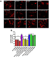
Figure 5. Helix-A nano blocks gp120-mediated neurite pruning
A. Cortical neurons were exposed to boiled gp120 (control) or to the indicated gp120s (all 5 nM) alone or in combination with Helix-A peptide or Helix-A nano (5 µM each) for 24 hr. Neurons were then fixed and stained for MAP2 as described in Materials and Methods. A. Representative confocal images of MAP2 positive (red) processes (Bar=20µm). B. Quantitative analysis of MAP2 positive processes after various conditions. Data are the mean ± SEM of three independent experiments (n=60 neurons per group per experiment) *p<0.01 vs control. No effect was observed with MSNs alone.
Figure 6
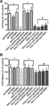
Figure 6. Helix-A nano prevents gp120-induced neuronal cell death
Cortical neurons were exposed to the indicated stimuli. Cell death was determined by (A) Hoechst/PI staining and (B) LDH 24 hr after gp120s. Data are the mean ± SEM of three separate experiments (n=200 neurons each group per experiment). *p<0.001 vs control; **p<0.01 vs gp120. #p<0.01 vs gp120. Helix-A nano blocked gp120-mediated cell death up to 96 hr whereas MSNs alone did not (data not shown).

Similar articles

Cited by

References

    1. Ahmed F, MacArthur L, De Bernardi MA, Mocchetti I. Retrograde and anterograde transport of HIV protein gp120 in the nervous system. Brain Behav Immun. 2009;23:355–364. - PMC - PubMed
    1. Altar CA, Cai N, Bliven T, Juhasz M, Conner JM, Acheson AL, Lindsay RM, Wiegand SJ. Anterograde transport of brain-derived neurotrophic factor and its role in the brain. Nature. 1997;389:856–860. - PubMed
    1. Avdoshina V, Biggio F, Palchik G, Campbell LA, Mocchetti I. Morphine induces the release of CCL5 from astrocytes: potential neuroprotective mechanism against the HIV protein gp120. Glia. 2010;58:1630–1639. - PMC - PubMed
    1. Avdoshina V, Fields JA, Castellano P, et al. The HIV protein gp120 alters mitochondrial dynamics in neurons. Neurotox Res. (in press) - PMC - PubMed
    1. Bachis A, Aden SA, Nosheny RL, Andrews PM, Mocchetti I. Axonal transport of human immunodeficiency virus type 1 envelope protein glycoprotein 120 is found in association with neuronal apoptosis. J Neurosci. 2006;26:6771–6780. - PMC - PubMed

Publication types

MeSH terms