

In vitro models of axon regeneration
- PMID: 26826447
- PMCID: PMC4963305
- DOI: 10.1016/j.expneurol.2016.01.020
In vitro models of axon regeneration
Abstract
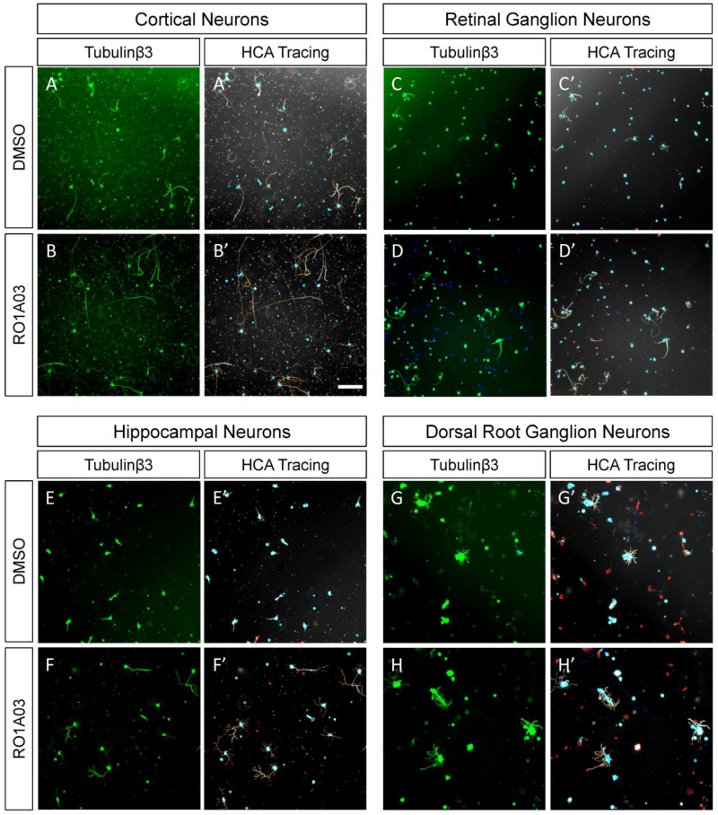
A variety of in vitro models have been developed to understand the mechanisms underlying the regenerative failure of central nervous system (CNS) axons, and to guide pre-clinical development of regeneration-promoting therapeutics. These range from single-cell based assays that typically focus on molecular mechanisms to organotypic assays that aim to recapitulate in vivo behavior. By utilizing a combination of models, researchers can balance the speed, convenience, and mechanistic resolution of simpler models with the biological relevance of more complex models. This review will discuss a number of models that have been used to build our understanding of the molecular mechanisms of CNS axon regeneration.
Keywords: Axon regeneration; Cell based assays; High content screening; In vitro models; Neurite outgrowth; Organotypic assays.
Copyright © 2016 Elsevier Inc. All rights reserved.
Figures
References
-
- Aguayo AJ, Dickson R, Trecarten J, Attiwell M, Bray GM, Richardson P. Ensheathment and myelination of regenerating PNS fibres by transplanted optic nerve glia. Neurosci. Lett. 1978;9:97–104. - PubMed
-
- Weinberg EL, Spencer PS. Studies on the control of myelinogenesis. 3. Signalling of oligodendrocyte myelination by regenerating peripheral axons. Brain Res. 1979;162:273–9. - PubMed
-
- Aguayo AJ, David S, Bray GM. Influences of the glial environment on the elongation of axons after injury: transplantation studies in adult rodents. J. Exp. Biol. 1981;95:231–40. - PubMed
-
- Richardson PM, Issa VM, Aguayo AJ. Regeneration of long spinal axons in the rat. J. Neurocytol. 1984;13:165–82. - PubMed
-
- Edelman GM. Surface modulation in cell recognition and cell growth. Science. 1976;192:218–26. - PubMed
Publication types
MeSH terms
Grants and funding
LinkOut - more resources
Full Text Sources
Other Literature Sources
Medical
