. 2016 Jan;8(1):127-46.
doi: 10.18632/aging.100879.

Age-related deficits in skeletal muscle recovery following disuse are associated with neuromuscular junction instability and ER stress, not impaired protein synthesis

Affiliations

Age-related deficits in skeletal muscle recovery following disuse are associated with neuromuscular junction instability and ER stress, not impaired protein synthesis

Leslie M Baehr et al. Aging (Albany NY). 2016 Jan.

Abstract

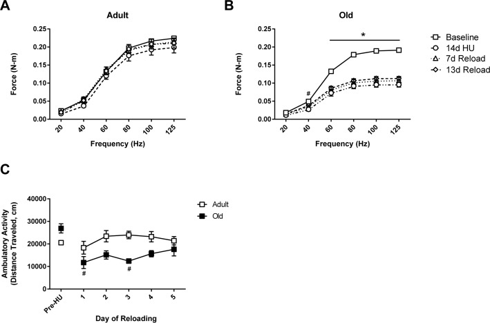
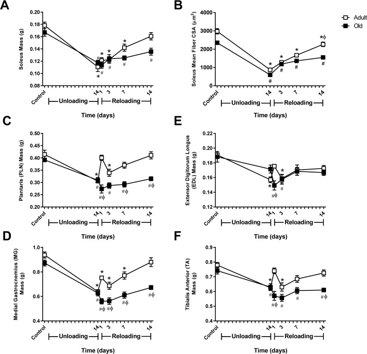
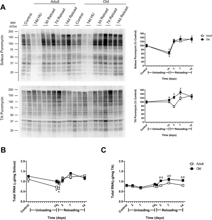
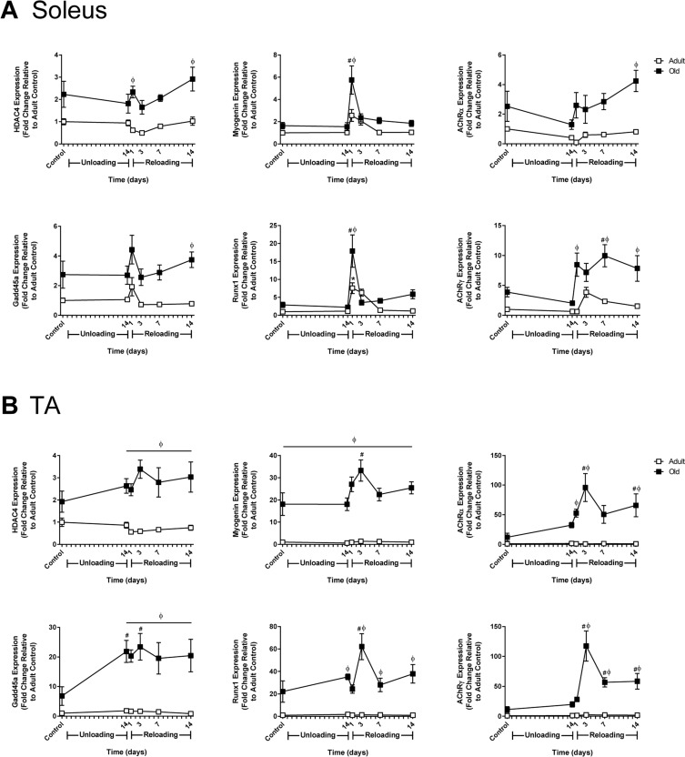
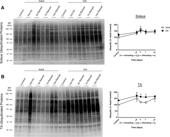
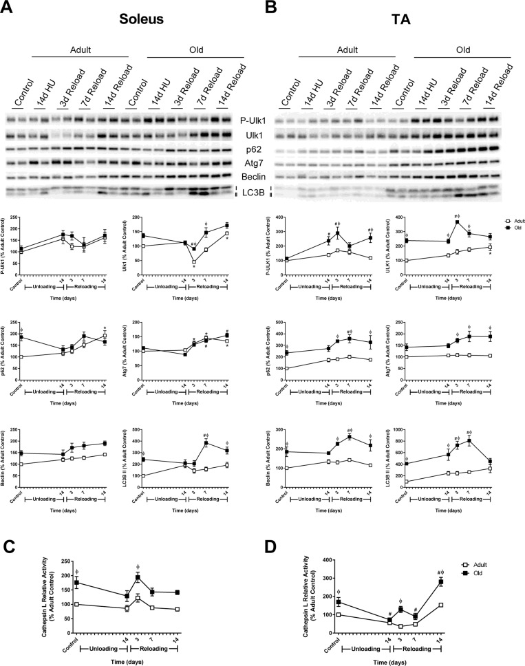
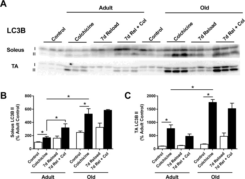
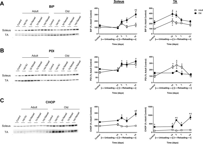
Age-related loss of muscle mass and strength can be accelerated by impaired recovery of muscle mass following a transient atrophic stimulus. The aim of this study was to identify the mechanisms underlying the attenuated recovery of muscle mass and strength in old rats following disuse-induced atrophy. Adult (9 month) and old (29 month) male F344BN rats underwent hindlimb unloading (HU) followed by reloading. HU induced significant atrophy of the hindlimb muscles in both adult (17-38%) and old (8-29%) rats, but only the adult rats exhibited full recovery of muscle mass and strength upon reloading. Upon reloading, total RNA and protein synthesis increased to a similar extent in adult and old muscles. At baseline and upon reloading, however, proteasome-mediated degradation was suppressed leading to an accumulation of ubiquitin-tagged proteins and p62. Further, ER stress, as measured by CHOP expression, was elevated at baseline and upon reloading in old rats. Analysis of mRNA expression revealed increases in HDAC4, Runx1, myogenin, Gadd45a, and the AChRs in old rats, suggesting neuromuscular junction instability/denervation. Collectively, our data suggests that with aging, impaired neuromuscular transmission and deficits in the proteostasis network contribute to defects in muscle fiber remodeling and functional recovery of muscle mass and strength.

Keywords: aging; anabolic resistance; autophagy; hindlimb unloading; ubiquitin proteasome system.


Conflict of interest statement

Conflict of interest statement

The authors declare no conflicts of interest.

Figures

Figure 1
Figure 1. Force output and activity measures in adult and old rats
In vivo isometric force production at frequencies ranging from 20-125 Hz was measured in (A) adult (9 mo) and (B) old (29 mo) rats prior to hindlimb unloading (HU) (baseline, open squares), after 14 days of HU (open circles), and then after 7 (open triangles) and 13 days of reloading (open diamonds). n=6/group. Values are mean ± SEM, *p<0.05 vs old baseline value, #p<0.05 for old 14d HU value vs old baseline value at 40 Hz. (C) Normal cage activity of adult (open squares, n=4) and old (filled squares, n=5) rats was recorded during the dark cycle for six days prior to HU and for the first five days of reloading following 14 days of HU. Values are mean ± SEM, #p<0.05 vs old control.
Figure 2
Figure 2. Comparison of muscle mass and soleus fiber cross-sectional area changes in adult and old rats subjected to hindlimb unloading (HU) and reloading
Adult (9 mo) and old (29 mo) male rats underwent HU for 14 days or underwent HU for 14 days and then were allowed to resume normal weight bearing activity for either 1, 3, 7, or 14 days. Average masses of the soleus (A), plantaris (C), medial gastrocnemius (D), extensor digitorum longus (E), and tibialis anterior (F) muscles of adult (open squares) and old (filled squares) rats at the various HU and reloading time points (n=6-7/group). (B) Changes in fiber cross-sectional area (CSA) were measured in the soleus of adult (open squares) and old (filled squares) rats after 14 days of HU and after 3, 7, and 14 days of reloading. Fiber CSA was determined from laminin-stained cross sections (n=5-6/group). Values are mean ± SEM, *p<0.05 vs adult control, #p<0.05 vs old control, φp<0.05 vs adult at same time point.
Figure 3
Figure 3. Effect of hindlimb unloading (HU) and reloading on muscle protein synthesis and total RNA in adult and old rats
(A) Using the SUnSET method, protein synthesis was measured in the soleus and tibialis anterior (TA) muscles of adult (9 mo, open squares) and old (29 mo, filled squares) rats after 14 days of HU and after 3, 7, and 14 days of reloading. Total protein, determined by stain-free imaging of the PVDF membrane, was used to normalize protein expression. Puromycin values are expressed as a percentage of each age-matched control group (n=6-7/group). Total RNA (μg/mg muscle) was measured in the soleus (B) and TA (C) muscles of adult (open squares) and old (filled squares) rats after 14 days of HU and after 1, 3, 7, and 14 days of reloading (n=6/group). Values are mean ± SEM, *p<0.05 vs adult control, #p<0.05 vs old control, φp<0.05 vs adult at same time point.
Figure 4
Figure 4. Effect of hindlimb unloading (HU) and reloading on gene expression markers of inactivity and/or denervation in adult and old rats
mRNA expression (fold change relative to adult control) of HDAC4, myogenin, Gadd45a, Runx1, and acetylcholine receptor subunits alpha (AChRα) and gamma (AChRγ) were measured in the soleus (A) and tibialis anterior (TA) (B) muscles of adult (9 mo, open squares) and old (29 mo, filled squares) rats after 14 days of HU and after 1, 3, 7, and 14 days of reloading (n=5-6/group). Values are mean ± SEM, *p<0.05 vs adult control, #p<0.05 vs old control, φp<0.05 vs adult at same time point.
Figure 5
Figure 5. Total ubiquitin levels in adult and old rats after hindlimb unloading (HU) and reloading
Representative Western blot and quantification of total ubiquitin levels in the soleus (A) and tibialis anterior (TA) (B) muscles of adult (9 mo, open squares) and old (29 mo, filled squares) rats after 14 days of HU and after 3, 7, and 14 days of reloading. Total protein, determined by stain-free imaging of the PVDF membrane, was used to normalize protein expression. Data are expressed as a percentage relative to the adult control group (n=5-6/group). Values are mean ± SEM, *p<0.05 vs adult control, #p<0.05 vs old control, φp<0.05 vs adult at same time point.
Figure 6
Figure 6. Changes in MuRF1 and MAFbx expression and proteasome activity in adult and old rats after hindlimb unloading (HU) and reloading
mRNA expression of MuRF1 and MAFbx was assessed by quantitative PCR in the soleus (A,B) and tibialis anterior (TA) (C,D) muscles of adult (9 mo, open squares) and old (29 mo, filled squares) rats after 14 days of HU and following 1, 3, 7, and 14 days of reloading. Gene expression was normalized to tissue weight. Data are expressed as a fold change relative to the adult control group. Proteolytic activity of the β1, β2, and β5 subunits of the 20S and 26S proteasome was measured in the soleus (E) and TA (F) muscles of adult (open squares) and old (filled squares) rats after 14 days of HU and after 3, 7, and 14 days of reloading. Data are expressed as a percentage relative to the activity of the adult control group for each subunit (n=4-6/group). Values are mean ± SEM, *p<0.05 vs adult control, #p<0.05 vs old control, φp<0.05 vs adult at same time point.
Figure 7
Figure 7. Autophagy-related changes in protein expression and cathepsin L activity during hindlimb unloading (HU) and reloading
Expression of the autophagy-related proteins phospho- and total Ulk1, p62, Atg7, Beclin, and LC3B-II was measured by Western blot and quantified after 14 days of HU and following 3, 7, and 14 days of reloading in the soleus (A) and tibialis anterior (TA) (B) muscles of adult (9 mo, open squares) and old (29 mo, filled squares) rats. Total protein, determined by stain-free imaging of the PVDF membrane, was used to normalize protein expression. Data are expressed as a percentage relative to the adult control group for each protein (n=4-6/group). Cathepsin L activity was measured by fluorometric assay after 14 days of HU and following 3, 7, and 14 days of reloading in the soleus (C) and TA (D) muscles of adult (open squares) and old (filled squares) rats. Data are expressed as a percentage relative to the activity of the adult control group (n=5-6/group). Values are mean ± SEM, *p<0.05 vs adult control, #p<0.05 vs old control, φp<0.05 vs adult at same time point.
Figure 8
Figure 8. Effect of age and reloading on autophagic flux and p62 protein accumulation using the autophagy inhibitor colchicine
Representative Western blots (A) and quantification of LC3B-II protein expression in the soleus (B) and tibialis anterior (TA) (C) muscles of control and 7 day reloaded adult (9 mo) and old (29 mo) rats treated with or without colchicine. Total protein, determined by stain-free imaging of the PVDF membrane, was used to normalize protein expression. Data are expressed as a percentage relative to the adult control group for each protein (n=3-6/group). Values are mean ± SEM, *p<0.05.
Figure 9
Figure 9. Markers of endoplasmic reticulum (ER) stress in adult and old rats during hindlimb unloading (HU) and reloading
Representative Western blots and quantification of ER stress markers BiP (A), PDI (B), and CHOP (C) in the soleus and tibialis anterior (TA) muscles of adult (9 mo, open squares) and old (29 mo, filled squares) rats following 14 days of HU and after 3, 7, and 14 days of reloading. Total protein, determined by stain-free imaging of the PVDF membrane, was used to normalize protein expression. Data are expressed as a percentage relative to the adult control group for each protein (n=4-6/group). Values are mean ± SEM, *p<0.05 vs adult control, #p<0.05 vs old control, φp<0.05 vs adult at same time point.

Similar articles

Cited by

References

    1. Marcell TJ. Sarcopenia: causes, consequences, and preventions. The journals of gerontology Series, A, Biological sciences and medical sciences. 2003;58:M911–916. - PubMed
    1. Lieffers JR, Bathe OF, Fassbender K, Winget M, Baracos VE. Sarcopenia is associated with postoperative infection and delayed recovery from colorectal cancer resection surgery. British journal of cancer. 2012;107:931–936. - PMC - PubMed
    1. Prado CMM, Baracos VE, McCargar LJ, Reiman T, Mourtzakis M, Tonkin K, Mackey JR, Koski S, Pituskin E, Sawyer MB. Sarcopenia as a Determinant of Chemotherapy Toxicity and Time to Tumor Progression in Metastatic Breast Cancer Patients Receiving Capecitabine Treatment. Clinical Cancer Research. 2009;15:2920–2926. - PubMed
    1. Montano-Loza AJ. Severe muscle depletion predicts postoperative length of stay but is not associated with survival after liver transplantation. Liver Transplantation. 2014;20:1424–1424. - PubMed
    1. Carey EJ. Sarcopenia in Solid Organ Transplantation. Nutrition in Clinical Practice. 2014;29:159–170. - PubMed

Publication types

MeSH terms

Substances