

Review
doi: 10.1016/j.matbio.2016.01.014.
Epub 2016 Jan 29.
Keloids: Animal models and pathologic equivalents to study tissue fibrosis
Affiliations
- PMID: 26827712
- PMCID: PMC4842112
- DOI: 10.1016/j.matbio.2016.01.014
Item in Clipboard
Review
Keloids: Animal models and pathologic equivalents to study tissue fibrosis
Matrix Biol.
2016 Apr.
Abstract
Animal models are crucial for the study of fibrosis. Keloids represent a unique type of fibrotic scarring that occurs only in humans, thus presenting a challenge for those studying the pathogenesis of this disease and its therapeutic options. Here, several animal models of fibrosis currently in use are described, emphasizing recent progress and highlighting encouraging challenges.
Keywords: Animal models; Hypertrophic scar; Keloids; Tissue fibrosis.
Copyright © 2016 International Society of Matrix Biology. Published by Elsevier B.V. All rights reserved.
Figures
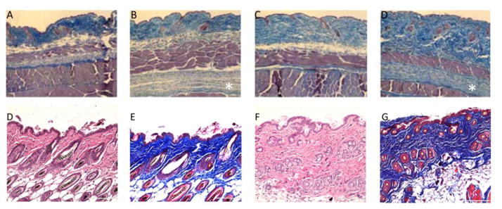
Hematoxylin & Eosin and Masson’s Trichrome staining of wild-type +/+ (A, C), Tsk/+ (B, D), PBS-treated (E,F) and bleomycin-treated (G, H) cutaneous sections. Genetic mutation in fibrillin-1 gene in the Tsk/+ mouse leads to thickened dermis with abundant collagen in both upper and lower dermis (B & D, sub-panniculus carnosus) compared to control (A & C). Daily administration of bleomycin elicits a strong inflammatory reaction in the host animal which, in turn, leads to increased dermal thickness and fibrosis (A & C) compared to control (B & D). Images of Tsk mouse reproduced from: Manne, J., Markova, M., Siracusa, L., Jimenez, S.A. “Collagen content in skin and internal organs of the Tight Skin Mouse: An animal model of scleroderma. Biochemistry Research International. Vol 2013, 1–8.
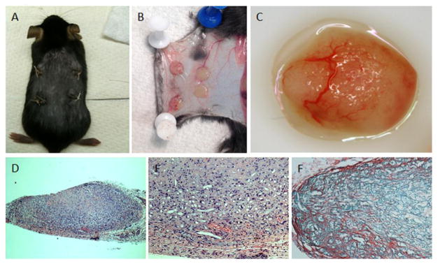
PLA scaffolds (B & C) are dynamically seeded and subcutaneously implanted into immune-deficient mice (A). Upon retrieval, the scaffolds demonstrate marked neovascularization from the host animal, and continued collagen production (D, E: H&E staining, F: Sirius red staining).
References
-
- Rubio-Rivas M, Royo C, Simeon CP, Corbella X, Fonollosa V. Mortality and survival in systemic sclerosis: systematic review and meta-analysis. Semin Arthritis Rheum. 2014;44:208–219. - PubMed
-
- Baxter RM, Crowell TP, McCrann ME, Frew EM, Gardner H. Analysis of the tight skin (Tsk1/+) mouse as a model for testing antifibrotic agents. Lab Invest. 2005;85:1199–1209. - PubMed
-
- Christner PJ, Peters J, Hawkins D, Siracusa LD, Jimenez SA. The tight skin 2 mouse. An animal model of scleroderma displaying cutaneous fibrosis and mononuclear cell infiltration. Arthritis Rheum. 1995;38:1791–1798. - PubMed
-
- Long KB, Li Z, Burgwin CM, Choe SG, Martyanov V, Sassi-Gaha S, Earl JP, Eutsey RA, Ahmed A, Ehrlich GD, Artlett CM, Whitfield ML, Blankenhorn EP. The Tsk2/+ mouse fibrotic phenotype is due to a gain-of-function mutation in the PIIINP segment of the Col3a1 gene. J Invest Dermatol. 2015;135:718–727. - PMC - PubMed
Publication types
MeSH terms
Substances
Grants and funding
LinkOut - more resources
Full Text Sources
Other Literature Sources
