. 2016 Mar:490:49-58.
doi: 10.1016/j.virol.2016.01.004. Epub 2016 Jan 30.

3B11-N, a monoclonal antibody against MERS-CoV, reduces lung pathology in rhesus monkeys following intratracheal inoculation of MERS-CoV Jordan-n3/2012

Affiliations

3B11-N, a monoclonal antibody against MERS-CoV, reduces lung pathology in rhesus monkeys following intratracheal inoculation of MERS-CoV Jordan-n3/2012

Reed F Johnson et al. Virology. 2016 Mar.

Abstract

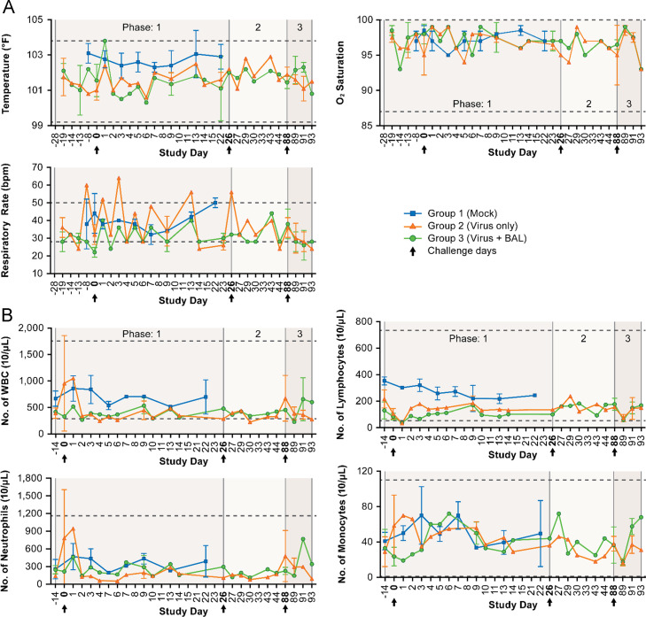
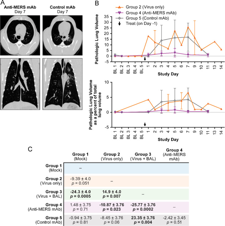
Middle East Respiratory Syndrome Coronavirus (MERS-CoV) was identified in 2012 as the causative agent of a severe, lethal respiratory disease occurring across several countries in the Middle East. To date there have been over 1600 laboratory confirmed cases of MERS-CoV in 26 countries with a case fatality rate of 36%. Given the endemic region, it is possible that MERS-CoV could spread during the annual Hajj pilgrimage, necessitating countermeasure development. In this report, we describe the clinical and radiographic changes of rhesus monkeys following infection with 5×10(6) PFU MERS-CoV Jordan-n3/2012. Two groups of NHPs were treated with either a human anti-MERS monoclonal antibody 3B11-N or E410-N, an anti-HIV antibody. MERS-CoV Jordan-n3/2012 infection resulted in quantifiable changes by computed tomography, but limited other clinical signs of disease. 3B11-N treated subjects developed significantly reduced lung pathology when compared to infected, untreated subjects, indicating that this antibody may be a suitable MERS-CoV treatment.

Keywords: Animal model, MERS; Antibody therapy; Human monoclonal antibody therapy; MERS-CoV; Respiratory syndrome.


Figures

Fig. 1.
Fig. 1
5 Groups of rhesus monkeys were challenged by IT infection with 5×10⁶ PFU of MERS-JOR or γ-irradiated virus as shown. Periodic CT and blood draws were performed; N represents the number of subjects from each group that underwent the indicated procedures. The variation in number of procedures performed on a given day was logistically determined based on blood withdrawal limits and accessibility to the CT, Each subject was given a complete physical exam prior to CT or blood withdrawal.
Fig. 2.
Fig. 2
Rhesus monkeys demonstrated limited clinical indicators of disease in response to infection with MERS-CoV Jordan-n3/2012. (A) Temperature, respiratory rate and peripheral oxygen saturation were within normal ranges for rhesus monkeys. (B) CBC/Diff data indicating limited response to MERS-CoV inoculation. WBC, lymphocyte, neutrophil and monocyte total counts were within normal ranges, although some changes are observed. Hashed lines indicate normal value ranges. Group 1 Mock (blue squares), Group 2 5×10⁶ PFU (orange triangles), and Group 3, Green circles (5×10⁶ PFU w/BAL). Arrows indicate challenge and re-challenge days. The shaded backgrounds represent the different phases of the experiment, initial challenge, followed by the 2nd and 3rd challenges. (For interpretation of the references to color in this figure legend, the reader is referred to the web version of this article.)
Fig. 3.
Fig. 3
MERS-CoV exposure of rhesus results in mild, but quantifiable lung pathology. (A) Lung volume quantification indicates mild, virus specific changes. Left panel is the total abnormal lung volume as quantified from CT data for Groups 1, 2, and 3. The data demonstrate that lung pathology is transient and re-challenge does not result in increased lung pathology. Right panel is the quantification of the CT as a percentage of total lung volume. Asterisks indicate days at which peak lung pathology was observed for individual subjects Group 1 Mock (blue squares), Group 2 5×10⁶ PFU (orange triangles), and Group 3, 5×10⁶ PFU w/BAL (green circles) Arrows indicate challenge and re-challenge days. The shaded backgrounds represent the different phases of the experiment, initial challenge, followed by the 2nd and 3rd challenges. (B) Representative CT of selected subjects from each group demonstrating peak lung pathology. (For interpretation of the references to color in this figure legend, the reader is referred to the web version of this article.)
Fig. 4
Fig. 4
(A) Representative CT of selected subjects from each group demonstrating peak lung pathology. (B) Comparison between 3B11-N (Group 4, purple inverted triangle) and 4E10-N (Group 5, gray diamond). Treatment with 3B11-N resulted in statistically significantly (p<0.0001) decreased lung pathology when compared to the untreated, infected group. Untreated, infected subjects (Group 2, orange triangle) were included for comparison. (C) Statistical comparisons between all groups which supports that BAL exacerbates disease. Days 0–14 for each group were analyzed by a linear mixed model. The summarized mean difference, standard error and P value are given in the table below. A P value of 0.01 was used to provide a stringent basis for significance. Comparisons with statistically significant differences are in bold. (For interpretation of the references to color in this figure legend, the reader is referred to the web version of this article.)

References

    1. Adney D.R., van Doremalen N., Brown V.R., Bushmaker T., Scott D., de Wit E., Bowen R.A., Munster V.J. Replication and shedding of MERS-CoV in upper respiratory tract of inoculated dromedary camels. Emerg. Infect. Dis. 2014;20:1999–2005. - PMC - PubMed
    1. Agrawal A.S., Garron T., Tao X., Peng B.H., Wakamiya M., Chan T.S., Couch R.B., Tseng C.T. Generation of a transgenic mouse model of Middle East respiratory syndrome coronavirus infection and disease. J. Virol. 2015;89:3659–3670. - PMC - PubMed
    1. Bossart K.N., Zhu Z., Middleton D., Klippel J., Crameri G., Bingham J., McEachern J.A., Green D., Hancock T.J., Chan Y.P., Hickey A.C., Dimitrov D.S., Wang L.F., Broder C.C. A neutralizing human monoclonal antibody protects against lethal disease in a new ferret model of acute nipah virus infection. PLoS Pathog. 2009;5:e1000642. - PMC - PubMed
    1. Channappanavar R., Fett C., Zhao J., Meyerholz D.K., Perlman S. Virus-specific memory CD8 T cells provide substantial protection from lethal severe acute respiratory syndrome coronavirus infection. J. Virol. 2014;88:11034–11044. - PMC - PubMed
    1. Channappanavar R., Zhao J., Perlman S. T cell-mediated immune response to respiratory coronaviruses. Immunol. Res. 2014;59:118–128. - PMC - PubMed

Publication types

MeSH terms