Review
. 2016 Jan 27;5(2):17.
doi: 10.3390/jcm5020017.

Role of EMT in Metastasis and Therapy Resistance

Affiliations
Review

Role of EMT in Metastasis and Therapy Resistance

Bethany N Smith et al. J Clin Med. .

Abstract

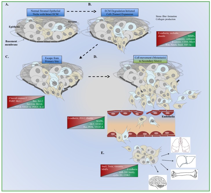
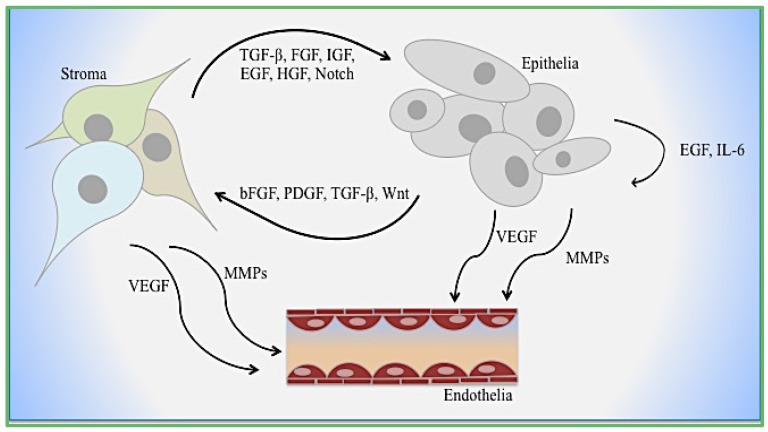
Epithelial-mesenchymal transition (EMT) is a complex molecular program that regulates changes in cell morphology and function during embryogenesis and tissue development. EMT also contributes to tumor progression and metastasis. Cells undergoing EMT expand out of and degrade the surrounding microenvironment to subsequently migrate from the primary site. The mesenchymal phenotype observed in fibroblasts is specifically important based on the expression of smooth muscle actin (α-SMA), fibroblast growth factor (FGF), fibroblast-specific protein-1 (FSP1), and collagen to enhance EMT. Although EMT is not completely dependent on EMT regulators such as Snail, Twist, and Zeb-1/-2, analysis of upstream signaling (i.e., TGF-β, EGF, Wnt) is necessary to understand tumor EMT more comprehensively. Tumor epithelial-fibroblast interactions that regulate tumor progression have been identified during prostate cancer. The cellular crosstalk is significant because these events influence therapy response and patient outcome. This review addresses how canonical EMT signals originating from prostate cancer fibroblasts contribute to tumor metastasis and recurrence after therapy.

Keywords: EMT; microenvironment; prostate cancer; stroma.


Figures

Figure 1
Figure 1
Summary of EMT and metastasus. (A) Normal interactions between cells and extracellular matrix (ECM) compobnents. Changes in canonical signaling and cellular function are the foundation of aberrant cell growth and expansion; (B) Increased expression of enzymes that degrade the ECM are involved in decreasing adhesion to the basement membrane; (C) Cells escape from the basement membrane as growth factor and cytokine signaling increases to accommodate changes in the microenvironment; (D) Cells intravasate and spread to the bloodstream as circulating tumor cells (CTC); (E) Extravasation at the secondary site promotes the formation of micrometastases and the re-induction of epithelial markers (MET). Sites of metastasis include the lungs, liver, bones, and brain.
Figure 2
Figure 2
Cellular interactions during EMT. Mechanisms of epithelial–stromal–endothelial crosstalk are depicted. Growth factors, cytokines, and enzymes function dynamically with and between each cell type.

Similar articles

Cited by

References

    1. Quail D.F., Joyce J.A. Microenvironmental regulation of tumor progression and metastasis. Nat. Med. 2013;19:1423–1437. doi: 10.1038/nm.3394. - DOI - PMC - PubMed
    1. Kalluri R., Weinberg R.A. The basics of epithelial-mesenchymal transition. J. Clin. Invest. 2009;119:1420–1428. doi: 10.1172/JCI39104. - DOI - PMC - PubMed
    1. Kovacic J.C., Mercader N., Torres M., Boehm M., Fuster V. Epithelial-to-mesenchymal and endothelial-to-mesenchymal transition. From: Cardiovascular Development to Disease. Circulation. 2012;125:1795–1808. doi: 10.1161/CIRCULATIONAHA.111.040352. - DOI - PMC - PubMed
    1. Whiteside T.L. The tumor microenvironment and its role in promoting tumor growth. Oncogene. 2008;27:5904–5912. doi: 10.1038/onc.2008.271. - DOI - PMC - PubMed
    1. Acloque H., Adams M.S., Fishwick K., Bronner-Fraser M., Nieto M.A. Epithelial mesenchymal transitions: The importance of changing cell state in development and disease. J. Clin. Invest. 2009;119:1438–1449. doi: 10.1172/JCI38019. - DOI - PMC - PubMed