. 2016 Feb 1;11(2):e0148184.
doi: 10.1371/journal.pone.0148184. eCollection 2016.

Neuropathogenicity of Two Saffold Virus Type 3 Isolates in Mouse Models

Affiliations

Neuropathogenicity of Two Saffold Virus Type 3 Isolates in Mouse Models

Osamu Kotani et al. PLoS One. .

Abstract

Objective: Saffold virus (SAFV), a picornavirus, is occasionally detected in children with acute flaccid paralysis, meningitis, and cerebellitis; however, the neuropathogenicity of SAFV remains undetermined.

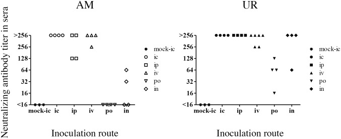
Methods: The virulence of two clinical isolates of SAFV type 3 (SAFV-3) obtained from a patient with aseptic meningitis (AM strain) and acute upper respiratory inflammation (UR strain) was analyzed in neonatal and young mice utilizing virological, pathological, and immunological methods.

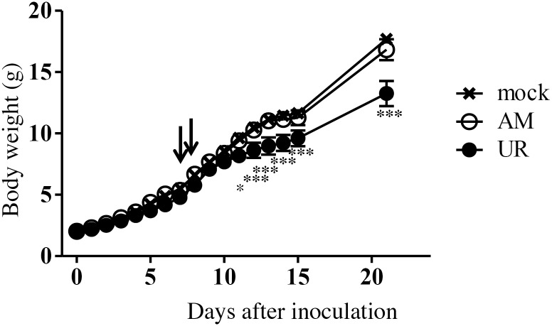
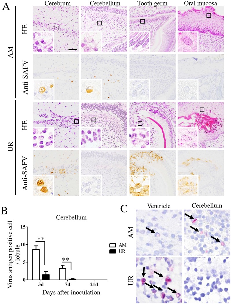
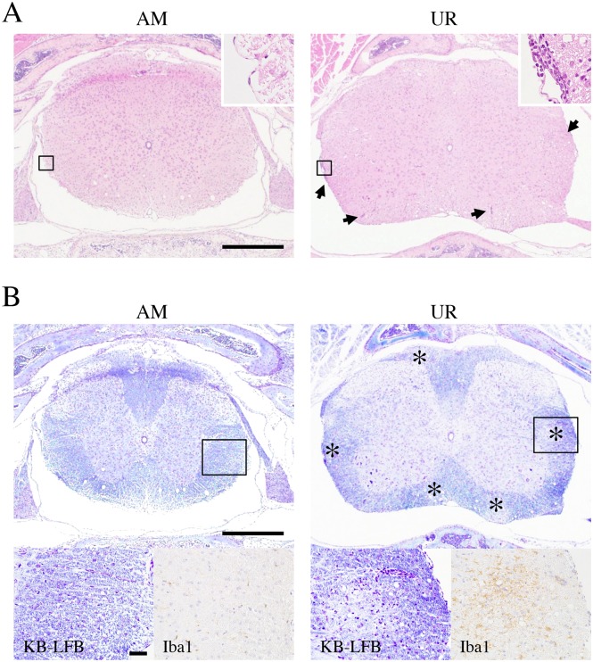
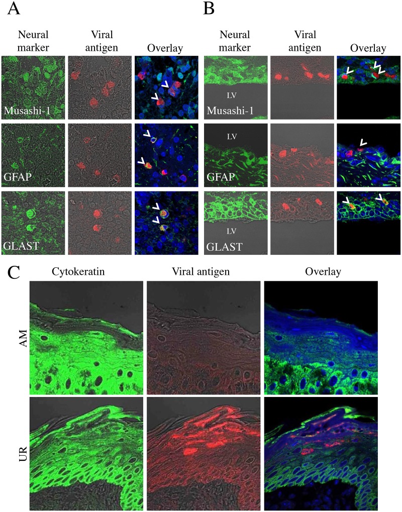
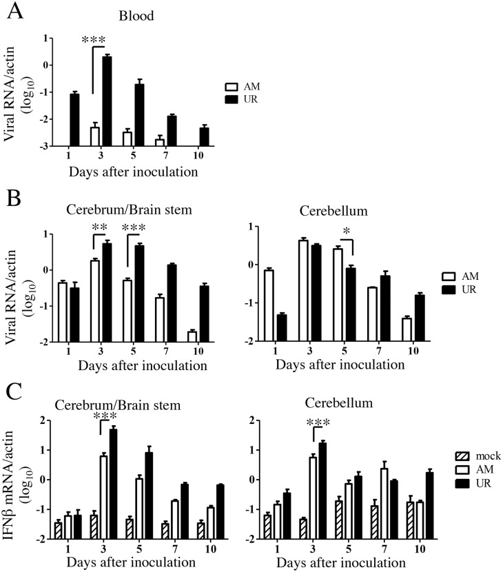
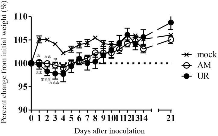
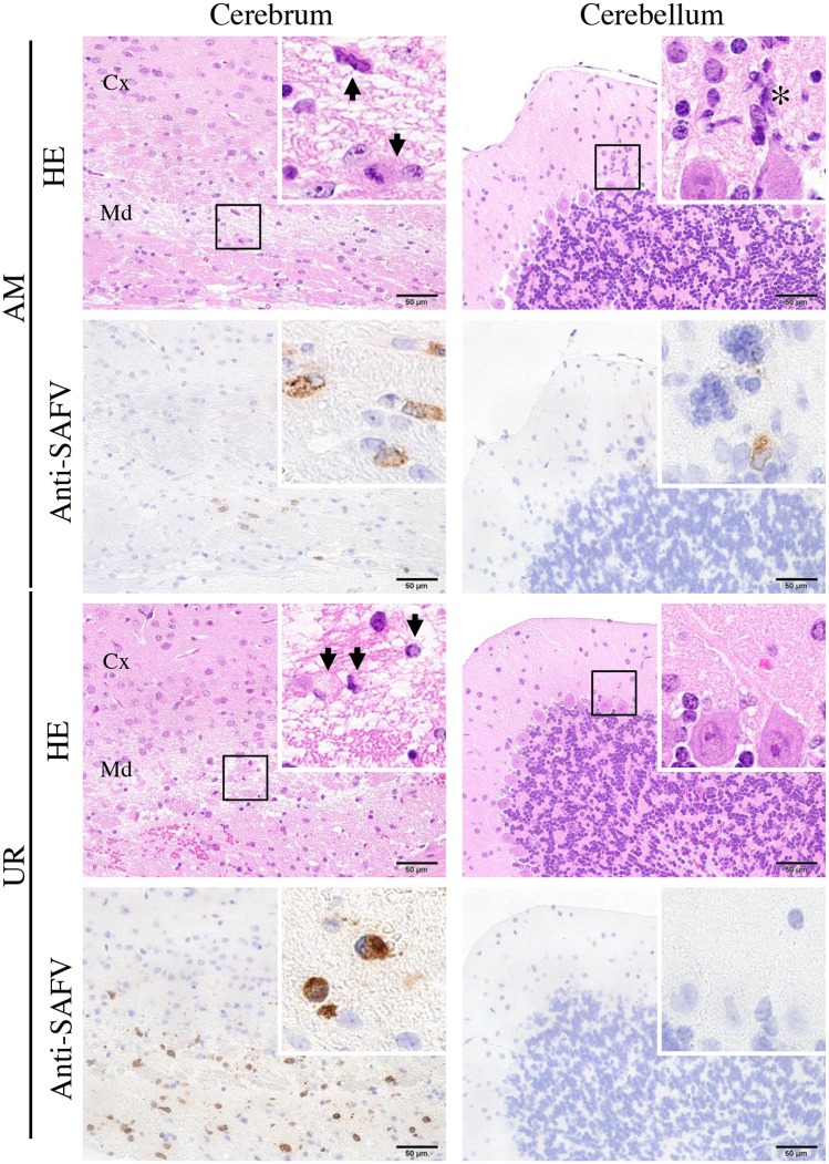
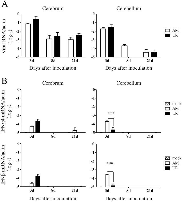
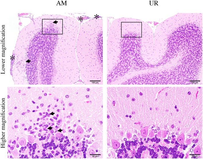
Results: The polyproteins of the strains differed in eight amino acids. Both clinical isolates were infective, exhibited neurotropism, and were mildly neurovirulent in neonatal ddY mice. Both strains pathologically infected neural progenitor cells and glial cells, but not large neurons, with the UR strain also infecting epithelial cells. UR infection resulted in longer inflammation in the brain and spinal cord because of demyelination, while the AM strain showed more infectivity in the cerebellum in neonatal ddY mice. Additionally, young BALB/c mice seroconverted following mucosal inoculation with the UR, but not the AM, strain.

Conclusions: Both SAFV-3 isolates had neurotropism and mild neurovirulence but showed different cell tropisms in both neonatal and young mouse models. This animal model has the potential to recapitulate the potential neuropathogenicity of SAFV-3.


Conflict of interest statement

Competing Interests: The authors have declared that no competing interests exist.

Figures

Fig 1
Fig 1. Clinical course of neonatal ddY mice after intracerebral inoculation with SAFV-3.
Within 24 h of birth, neonatal ddY mice were inoculated intracerebrally with 104 CCID50 (cell culture infectious dose) of the aseptic meningitis (AM) or upper respiratory (UR) strains of SAFV-3. Animals were observed for clinical manifestations, and body weight was measured daily for 15 days and on Day 21 (n = 3–4 mice per group). Two of the four AM-inoculated mice showed mild neurological signs, such as rolling and ataxia, on Days 7 and 8 post-inoculation (p.i.) (arrows), but the animals rapidly recovered. UR-inoculated mice showed significantly less weight gain than mock-infected control mice. All animals survived the inoculation. (*P < 0.05 and ***P < 0.001; one-way ANOVA). The data in the figure are representative of two experiments with similar results.
Fig 2
Fig 2. Histopathological examination of neonatal ddY mice inoculated intracerebrally with SAFV-3.
Within 24 h of birth, neonatal ddY mice were inoculated intracerebrally with 104 CCID50 (cell culture infectious dose) of the aseptic meningitis (AM) or upper respiratory (UR) strains of SAFV-3. (A) Representative histopathological findings of viral infection in neonatal mice 3 days post-inoculation (p.i.) are shown (n = 3–4 mice per group). Hematoxylin and eosin staining (H&E) and immunohistochemical analysis with an anti-SAFV-3 antibody (anti-SAFV). Bar, 50 μm. Mild histopathological changes were associated with viral antigen-positive cells (brown) in AM-inoculated neonatal mice. Degenerated and necrotic cells and a mild inflammatory infiltrate were observed around the lateral ventricle, in the tooth germ, and in the oral mucosa of UR-inoculated neonatal mice. Viral antigen-positive cells were present in the lesion. The cytoplasm of degenerated cells (with condensed nuclei) in the cerebrum and tooth germ was positive for viral antigens (insets). Meanwhile, the oral mucosa of UR-inoculated mice appeared histopathologically normal, but was positive for viral antigens (inset). (B) Number of viral antigen-positive cells in the cerebellum. (**P < 0.01; unpaired t-test). (C) In situ hybridization to visualize viral RNA in the brain of a neonatal mouse on Day 3 p.i. Viral RNA positive signals (red; arrows). Viral RNA was observed in viral antigen-positive tissue. Original magnification, 400× (A), and 1,000× (insets in A and C).
Fig 3
Fig 3. Demyelination in the spinal cord of neonatal ddY mice after intracerebral inoculation with SAFV-3.
Within 24 h of birth, neonatal ddY mice were inoculated intracerebrally with 104 CCID50 (cell culture infectious dose) of the aseptic meningitis (AM) or upper respiratory (UR) strains of SAFV-3. (A) Representative histopathological findings of the spinal cord in neonatal mice 21 days post-inoculation (p.i.) are shown. Hematoxylin and eosin staining (H&E) Bar, 500 μm. Meningitis and mild perivascular cuffing (arrows, upper right panels; box, low magnification) are observed in the white matter of the UR-inoculated mice on day 21 p.i. (B) Klüver-Barrera Luxol fast blue staining (KB-LFB) (upper panels; low magnification, lower left panels; box of upper panels) and immunohistochemical analysis with an anti-Iba1 antibody (lower right panels). Scattered demyelinations (asterisks) with inflammatory infiltrations were observed in the white matter of UR-inoculated mice. Infiltrated cells were Iba1 positive microglia (brown, lower right panels). Upper bar, 500 μm; lower bar, 50 μm. Upper panels, original magnification (50×); lower panels, 400×.
Fig 4
Fig 4. Identification of SAFV-3-infected cells in the neonatal mouse brain.
Within 24 h of birth, neonatal ddY mice were inoculated intracerebrally with 104 CCID50 (cell culture infectious dose) of the aseptic meningitis (AM) or upper respiratory (UR) strains of SAFV-3. Double immunofluorescent images showing viral antigens (red) and markers (green) for Musashi-1+ neural progenitor cells, GFAP+ astrocytes, and GLAST+ radial astrocytes, in the brains of mice on Day 3 post-inoculation (p.i.). (A) Viral antigen-positive cells in the cerebellum of AM-inoculated mice were identified as Musashi-1+ neural progenitor cells, and GFAP+ and GLAST+ radial astrocytes (aka Bergmann glia) (B) By contrast, Musashi-1+ neural progenitor cells (aka neuroepithelial cells), and GFAP+ and GLAST+ radial astrocytes in the ventricular zone of UR-inoculated mice were positive for viral antigens. Bar, 20 μm. (C) Viral antigen-positive cells in the oral mucosa of UR-inoculated mice were cytokeratin-positive epithelial cells. Arrows, viral antigen-positive and neural marker-positive cells (A, B). LV, lateral ventricle (B). Original magnification, 600×.
Fig 5
Fig 5. Viral replication and type 1 interferon expression in SAFV-3-inoculated neonatal ddY mice.
Within 24 h of birth, neonatal ddY mice were inoculated intracerebrally with 104 CCID50 (cell culture infectious dose) of the aseptic meningitis (AM) or upper respiratory (UR) strains of SAFV-3. The number of viral RNA copies was measured in the blood (A) and cerebrum/brain stem and cerebellum (B) of each mouse on Days 1, 3, 5, 7, and 10 post-inoculation (p.i.) (n = 4 mice per group for each day). The number of viral RNA copies is expressed relative to the number of mouse beta-actin RNA copies. Viral RNA levels in the blood and brain peaked at 3 to 5 Days p.i. in mice inoculated with AM and UR strains (*P < 0.05, **P < 0.01, and ***P < 0.001; unpaired t-test). (C) Real-time reverse transcriptase (RT)-PCR quantification of interferon beta (IFN-β) mRNA expression in the brains of SAFV-inoculated neonatal mice (n = 3–4 mice per group). The number of IFN-β RNA copies is expressed relative to the number of mouse beta-actin RNA copies. On Day 3 p.i., UR-inoculated mice showed greater expression of IFN-β than AM-inoculated mice (*P < 0.05 and ***P < 0.001; one-way ANOVA).
Fig 6
Fig 6. Body weight changes in young BALB/c mice after intracerebral inoculation with SAFV-3.
BALB/c mice were inoculated intracerebrally with 104 CCID50 (cell culture infectious dose) of the aseptic meningitis (AM) and upper respiratory (UR) strains of SAFV-3. Body weight was measured for 21 days post-inoculation (p.i.) (n = 6). The dotted line represents 100%. (*P < 0.05, **P < 0.01, and ***P < 0.001; unpaired t-test). Body weight of mice inoculated with either of the two SAFV-3 strains decreased during the early phase of infection. The figure shows representative data from two experiments with similar results.
Fig 7
Fig 7. Histopathological examination of brains from young BALB/c mice after intracerebral inoculation with SAFV-3.
On Day 3 post-inoculation (p.i.), brains were obtained from BALB/c mice after intracerebral inoculation with 104 CCID50 (cell culture infectious dose) of the aseptic meningitis (AM) and upper respiratory (UR) strains of SAFV-3. Hematoxylin and eosin (H&E) staining and immunohistochemical analysis with an anti-SAFV-3 antibody (anti-SAFV). Bar, 50 μm. Nerve cells were degenerated, with eccentric nuclei (arrows), and the cerebral medulla was positive for viral antigens (insets, left panels). Several viral antigen-positive cells were seen in the lesions in both AM- and UR-inoculated mice. Nerve cells in the cerebral cortex were negative for viral antigen. Focal inflammatory infiltration (asterisk) by viral antigen-positive cells was observed in the molecular layer of the cerebellum of AM-inoculated mice, but not in that of UR-inoculated mice (insets, right panels). Purkinje cells were negative for viral antigens. Cx, Cortex; Md, Medulla. Original magnification, 400×; insets, 1,000×.
Fig 8
Fig 8. Viral replication and type 1 interferon expression in the brains of SAFV-3-inoculated young BALB/c mice.
BALB/c mice were inoculated intracerebrally with 104 CCID50 (cell culture infectious dose) of the aseptic meningitis (AM) and upper respiratory (UR) strains of SAFV-3. (A) After inoculation, the number of viral RNA copies was measured in the cerebrum and cerebellum of each mouse on Days 3, 8, and 21 p.i. (n = 3 mice per group for each day). There was no difference in the number of viral RNA copies between the AM- and UR-inoculated groups (unpaired t-test). (B) Quantification of type 1 interferon (IFN), IFN-α4, and IFN-β mRNA expression in the brains of SAFV-3-inoculated mice on Days 3, 8, and 21 p.i. (n = 3 mice per group for each day). Number of viral IFN-α4 and IFN-β RNA copies relative to that of mouse beta-actin copies (***P < 0.001; one-way ANOVA). On Day 3, the expression of IFN-α4 and IFN-β in the cerebellum of AM-inoculated mice was higher than that in UR-inoculated mice.
Fig 9
Fig 9. Inflammatory infiltration in the cerebellum of SAFV-3-inoculated young BALB/c mice.
BALB/c mice were inoculated intracerebrally with 104 CCID50 (cell culture infectious dose) of the aseptic meningitis (AM) and upper respiratory (UR) strains of Saffold virus type 3 (SAFV-3). Hematoxylin and eosin (H&E) staining. Bar, 100 μm (upper panels) and 20 μm (lower panels). Histopathological findings in the cerebellum on Day 8 p.i. Inflammatory cells were observed in the cerebellar cortex (arrows, low magnification) and meninges (asterisk) of AM-inoculated mice (left panels), but not in those of UR-inoculated mice (right panels). Mononuclear cells and rod-shaped cells (arrows, high magnification) were observed in the cortex and meninges of AM-inoculated, but not UR-inoculated, mice (lower panels). Purkinje cells were present in the lesion. The figure shows representative data from two experiments with similar results. Original magnification: upper panels, 200×, and lower panels, 1,000×.
Fig 10
Fig 10. Titers of neutralizing antibodies against SAFV-3 in sera from young BALB/c mice.
On Day 21 post-inoculation, sera were obtained from mice inoculated intracerebrally (ic), intraperitoneally (ip), intravenously (iv), orally (po), or intranasally (in) with the aseptic meningitis (AM) or upper respiratory (UR) strains of SAFV-3 (n = 4 or 6 mice per group). All mice inoculated with the UR strain, but not those inoculated with the AM strain, via the oral or intranasal routes, generated neutralizing antibodies.

Similar articles

Cited by

References

    1. Jones MS, Lukashov VV, Ganac RD, Schnurr DP. Discovery of a novel human picornavirus in a stool sample from a pediatric patient presenting with fever of unknown origin. J Clin Microbiol. 2007;45(7):2144–50. Epub 2007/04/27. 10.1128/JCM.00174-07 - DOI - PMC - PubMed
    1. Dickinson L, Griffiths AJ. The pathogenesis of experimental infections with encephalomyocarditis (EMC) virus. Br J Exp Pathol. 1966;47(1):35–44. Epub 1966/02/01. - PMC - PubMed
    1. Craighead JE, McLane MF. Diabetes mellitus: induction in mice by encephalomyocarditis virus. Science. 1968;162(3856):913–4. Epub 1968/11/22. . - PubMed
    1. Dal Canto MC, Lipton HL. Multiple sclerosis. Animal model:Theiler's virus infection in mice. Am J Pathol. 1977;88(2):497–500. Epub 1977/08/01. - PMC - PubMed
    1. Yoon JW, McClintock PR, Onodera T, Notkins AL. Virus-induced diabetes mellitus. XVIII. Inhibition by a nondiabetogenic variant of encephalomyocarditis virus. J Exp Med. 1980;152(4):878–92. Epub 1980/10/01. - PMC - PubMed

Publication types

MeSH terms

LinkOut - more resources