. 2016 Feb 1;11(2):e0146792.
doi: 10.1371/journal.pone.0146792. eCollection 2016.

CPEB and miR-15/16 Co-Regulate Translation of Cyclin E1 mRNA during Xenopus Oocyte Maturation

Affiliations

CPEB and miR-15/16 Co-Regulate Translation of Cyclin E1 mRNA during Xenopus Oocyte Maturation

Ania Wilczynska et al. PLoS One. .

Abstract

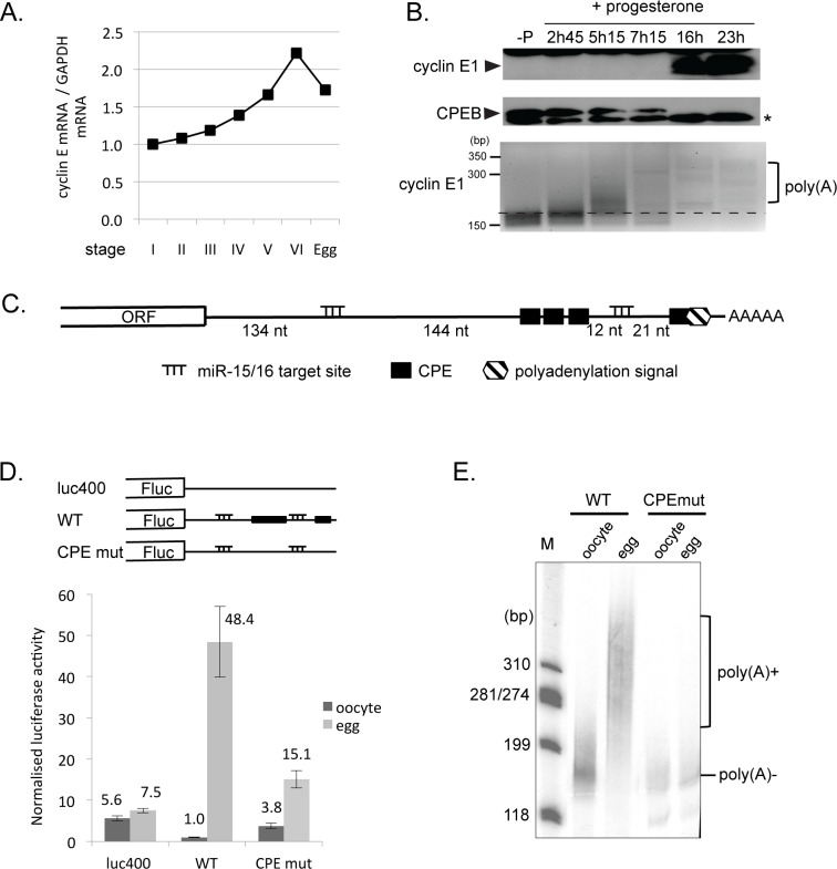
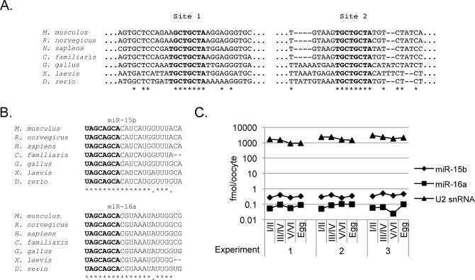
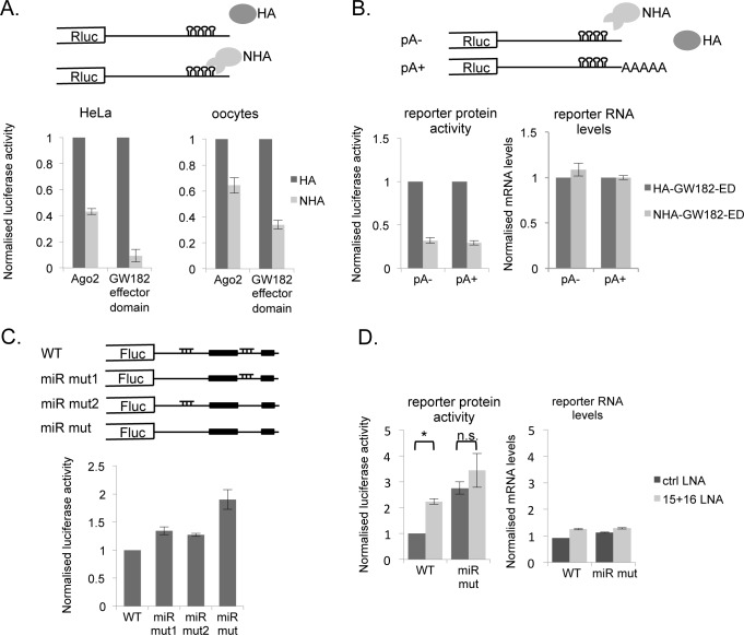
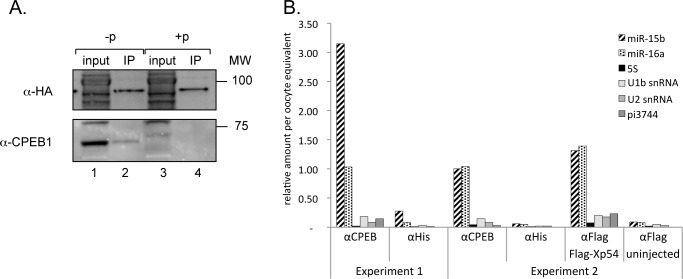
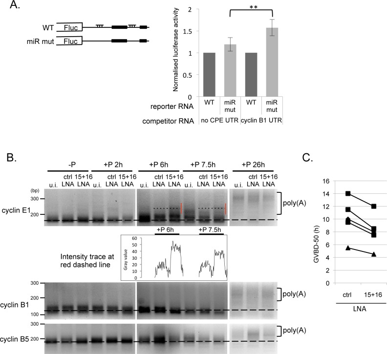
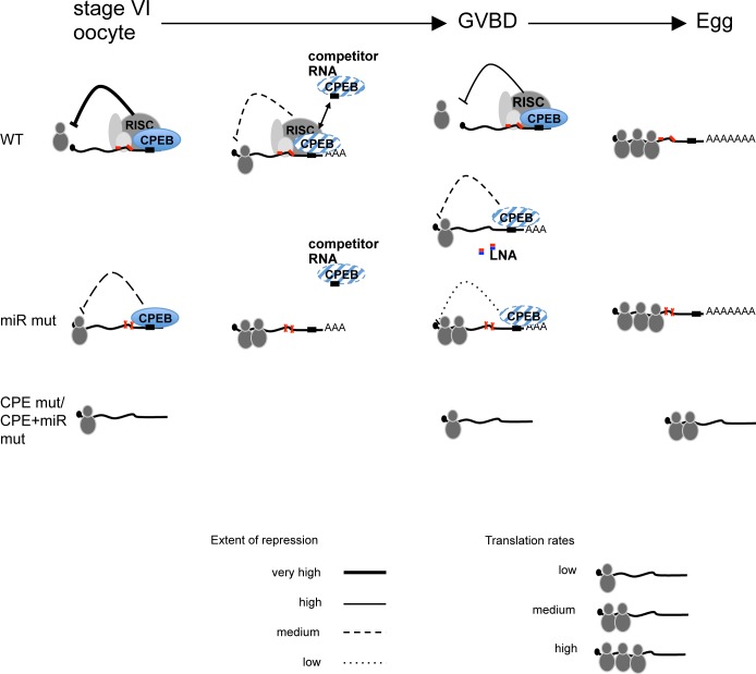
Cell cycle transitions spanning meiotic maturation of the Xenopus oocyte and early embryogenesis are tightly regulated at the level of stored inactive maternal mRNA. We investigated here the translational control of cyclin E1, required for metaphase II arrest of the unfertilised egg and the initiation of S phase in the early embryo. We show that the cyclin E1 mRNA is regulated by both cytoplasmic polyadenylation elements (CPEs) and two miR-15/16 target sites within its 3'UTR. Moreover, we provide evidence that maternal miR-15/16 microRNAs co-immunoprecipitate with CPE-binding protein (CPEB), and that CPEB interacts with the RISC component Ago2. Experiments using competitor RNA and mutated cyclin E1 3'UTRs suggest cooperation of the regulatory elements to sustain repression of the cyclin E1 mRNA during early stages of maturation when CPEB becomes limiting and cytoplasmic polyadenylation of repressed mRNAs begins. Importantly, injection of anti-miR-15/16 LNA results in the early polyadenylation of endogenous cyclin E1 mRNA during meiotic maturation, and an acceleration of GVBD, altogether strongly suggesting that the proximal CPEB and miRNP complexes act to mutually stabilise each other. We conclude that miR-15/16 and CPEB co-regulate cyclin E1 mRNA. This is the first demonstration of the co-operation of these two pathways.


Conflict of interest statement

Competing Interests: The authors have declared that no competing interests exist.

Figures

Fig 1
Fig 1. Translation of cyclin E1 during oocyte maturation is CPE-dependent.
A. Cyclin E1 mRNA is present throughout oogenesis. RT-qPCR was carried out on total RNA extracted from staged oocytes and eggs. The graph represents the relative amount of cyclin E1 mRNA compared to GAPDH mRNA, with values of each transcript set to 1 for stage I. Representative of 5 progesterone maturation experiments. B. Cyclin E1 protein is not expressed until late in oocyte maturation following the polyadenylation of its mRNA. Stage VI oocytes were incubated with progesterone for the indicated times and the corresponding lysates were analysed by Western blot using cyclin E1 and CPEB antibodies (upper panels). RNA extracted from these oocytes was subjected to RNA-ligation-coupled PCR poly(A) analysis (lower panel). *—non-specific band. C. The cyclin E1 3’UTR contains a cluster of three CPE sequences and an additional one overlapping the hexanucleotide as well as two putative miR-15/16 target sites as indicated. Not to scale. D. CPE sequences repress translation in immature oocytes, and activate translation in eggs. Schematic representation of reporters used. Firefly luciferase reporters containing a control 3’UTR (luc400), the wild-type cyclin E1 3’UTR (WT) or the 3’UTR with mutations in the CPEs (CPE mut) were injected into stage VI oocytes. Renilla luciferase reporter mRNA was co-injected as an internal control. Oocytes were incubated for 24h with or without progesterone and both sets were assayed for luciferase expression. Firefly luciferase levels are expressed as a ratio to Renilla internal control. E. CPE sequences in the cyclin E1 3’UTR direct polyadenylation during oocyte maturation. Radiolabelled RNAs representing the WT 3’-terminal 180 nt of the cyclin E1 3’UTR and the same fragment with mutated CPE sequences were injected into oocytes, and maturation was induced by progesterone. RNA extracted from untreated oocytes and progesterone-matured eggs was analysed by denaturing gel electrophoresis and autoradiography.
Fig 2
Fig 2. Mature forms of miR-15 and miR-16 are present in Xenopus oocytes.
A. Alignment of putative miR-15/16 seed binding sites across vertebrate cyclin E1 3’UTRs. B. Alignment of vertebrate miR-15b and miR-16a reveals nearly perfect conservation of the two miRNAs, with the seed sequence highlighted in bold. C. miR-15b and 16a levels do not undergo significant changes during oogenesis and oocyte maturation. Levels of the two microRNAs were verified by qPCR alongside the control U2 snRNA. The graph represents absolute quantities throughout oogenesis in three independent biological samples.
Fig 3
Fig 3. The miRISC as well as the putative miR-15/16 target sites in the cyclin E1 3’UTR are functional.
A. Tethering of either Ago2 or the effector domain of GW182 represses translation of a reporter in mammalian cells as well as in Xenopus oocytes. Schematic representation of reporters used. mRNAs encoding lambda-N peptide with an HA-tag or an HA-tag only, fused to GW182 effector domain or Ago2 were either injected into oocytes for 24 h prior to injection of Renilla luciferase reporter mRNA containing 3’UTR Box B sites (Rluc-BoxB) or transfected into HeLa cells 24 h prior to transfection of plasmids encoding the reporter genes. An mRNA/plasmid encoding Firefly luciferase was co-injected/co-transfected as an internal control. After a 6 h incubation, cells were harvested and reporter protein expression was assessed. The experiment was repeated 3 times with similar results. B. Repression of a reporter mRNA tethered to the effector domain of GW182 is independent of a poly(A) tail. Schematic representation of reporters used. The experiment was performed in Xenopus oocytes as in A, using either non-adenylated (pA-) or in vitro polyadenylated (pA+) RNA. RNA was assessed from a pool of 50 injected oocytes. C. The two miR-15/16 sites co-operate in repressing translation of the cyclin E1 3’UTR. Luciferase reporters containing the wild-type cyclin E1 3’UTR (WT), mutations in the first miR target site (miR mut1), the second target site (miR mut2) or both (miR mut) were injected into stage VI oocytes. Renilla luciferase mRNA was co-injected as an internal control. Firefly luciferase levels are expressed as a ratio to Renilla internal control. The graph displays the results for a representative experiment. D. The miR-15/16 target sites are active in a degradation-independent manner. Luciferase reporters containing the wild-type cyclin E1 3’UTR (WT) or the cyclin E1 miR mut 3’UTR were injected into oocytes (see C for further details), in the presence of co-injected control or miR-15/16 LNAs, as indicated. * Student t-test P<0.01. RNA extracted from injected oocytes was subjected to reverse transcription and quantitative real-time PCR to assess RNA levels, which are expressed as a ratio of firefly to Renilla luciferase. The graph displays the results for a representative experiment.
Fig 4
Fig 4. CPEB complexes and miRISC cooperate in the regulation of translational activation of cyclin E1 during oocyte maturation.
A. Components of the RISC complex can associate with the translational silencing complex in immature oocytes (-p). Oocytes were injected with HA-Ago2 mRNA, and the resulting lysates were subjected to immunoprecipitation with anti-HA antibodies. Input represents 5% of the immunoprecipitated fractions. Western blotting was performed with anti-HA or -CPEB1 antibodies and visualised by ECL.–p–no progesterone, immature oocytes; +p–oocytes matured with progesterone. B. miRNAs co-immunoprecipitate with CPEB complexes. Lysates from uninjected oocytes or oocytes expressing a FLAG-tagged Xp54 were used for immunoprecipitation using a monoclonal anti-CPEB antibody, an isotype-matched control (His), or in the case of FLAG-Xp54 expressing oocytes and uninjected control (“-“), an anti-FLAG antibody (FLAG). RNA was isolated and real-time RT-PCR performed for the miRNAs and small RNAs indicated. Relative quantities were normalised per oocyte input. For plotting purposes all enrichment values were scaled to the enrichment of miR-15 in the CPEB immunoprecipitate of Experiment 2.
Fig 5
Fig 5. Inhibition of miR-15/16 causes premature polyadenylation of cyclin E1 mRNA and acceleration of meiotic maturation.
A. Injection of a molar excess of CPE-containing RNA competitor enhances the effect of miR site mutations. 500 fmol of either a control 85 nt RNA not containing any CPE sequences (no CPE UTR) or a 65 nt 3’–terminal sequence of the cyclin B1 3’UTR (cyclin B1 UTR) were injected into stage VI oocytes. After an overnight incubation, oocytes were re-injected with either the WT or miR mut Firefly reporter constructs and the Renilla control RNA. The oocytes were lysed and assayed for luciferase after 6 h. The graph represents and average of 3 experiments with the WT reporter normalised to 1 in each case. (** P<0.01, 2-tailed paired t-test). B. Uninjected stage VI oocytes (u.i) or oocytes injected with control LNA (ctrl LNA) or a mixture of anti-miR-15 and anti-miR-16 LNAs (15+16 LNA) were incubated overnight, following which they were stimulated with progesterone. Samples were taken at indicated times. Extracted RNA was subjected to RNA-ligation coupled PCR polyadenylation analysis using primers for indicated mRNAs. Vertical red line indicates early polyadenylation in 15+16 LNA injected sample. Panel shows a representative experiment of 4. Graph underneath cyclin E panel depicts ImageJ profile plot taken along the dotted line. C. 100–150 stage VI oocytes were injected with either control LNA (ctrl LNA) or anti-miR-15 and anti-miR-16 LNAs (15+16 LNA) and incubated overnight and then treated with progesterone. GVBD was scored by the appearance of a white spot on the animal pole of the oocyte. The time required for 50% of the oocytes to achieve GVBD (GVBD-50) is plotted for 5 independent experiments.
Fig 6
Fig 6. Schematic summary of results and model of action of CPE- and microRNA-binding sites in cyclin E1 mRNA during meiotic maturation.
In the immature oocyte, the presence of CPE sequences on maternal mRNAs stabilises the association of miRISC with the target mRNA to cause augmented translational repression (WT vs. miR mut). In the absence of either the CPE sequence alone or both CPEs and miRNA target sites, the mRNA cannot bind the CPEB RNP nor RISC and is therefore not repressed. In the presence of RNAs competing for CPEB, miRISC acts to reinforce the interaction of the limited CPEB protein with the mRNA resulting in lower translation rates compared to the miR mut mRNA which does not bind miRISC. During GVBD, when the degradation of CPEB begins, once again, co-association of CPEB and miRISC with an mRNA stabilises both interactions and delays polyadenylation and translational activation. Mutation of the miRNA target sites or inhibition of miR-15/16 allows for early polyadenylation and activation of the transcript. In our model, we have not distinguished the functionally similar but temporally distinct roles of CPEB1 and CPEB4 [72].

References

    1. Minshall N, Walker J, Dale M, Standart N. Dual roles of p82, the clam CPEB homolog, in cytoplasmic polyadenylation and translational masking. RNA. 1999;5:27–38. - PMC - PubMed
    1. de Moor C, Richter JD. Cytoplasmic polyadenylation elements mediate masking and unmasking of cyclin B1 mRNA. EMBO J. 1999;18:2294–303. - PMC - PubMed
    1. Kamenska A, Simpson C, Standart N. eIF4E-binding proteins: New factors, new locations, new roles. Bioch Soc Trans. 2014;42:1238–45. - PubMed
    1. Richter JD. CPEB: a life in translation. Trends Biochem Sci. 2007;32:279–85. - PubMed
    1. Minshall N, Reiter M-H, Weil D, Standart N. CPEB interacts with an ovary-specific eIF4E and 4E-T in early Xenopus oocytes. J Biol Chem. 2007;282:37389–401. - PubMed

Publication types

MeSH terms