. 2016 Mar;34(3):334-8.
doi: 10.1038/nbt.3469. Epub 2016 Feb 1.

A dual AAV system enables the Cas9-mediated correction of a metabolic liver disease in newborn mice

Affiliations

A dual AAV system enables the Cas9-mediated correction of a metabolic liver disease in newborn mice

Yang Yang et al. Nat Biotechnol. 2016 Mar.

Abstract

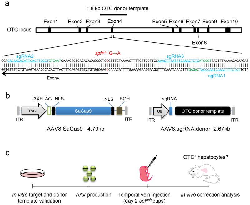
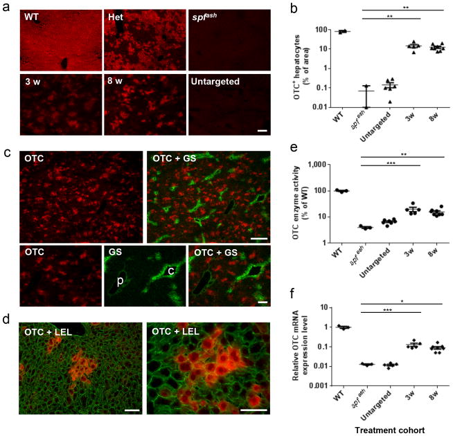
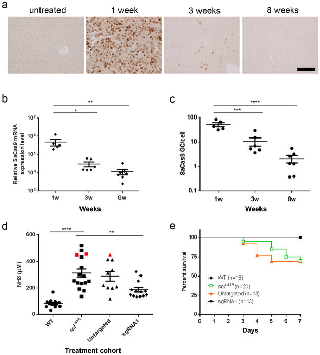
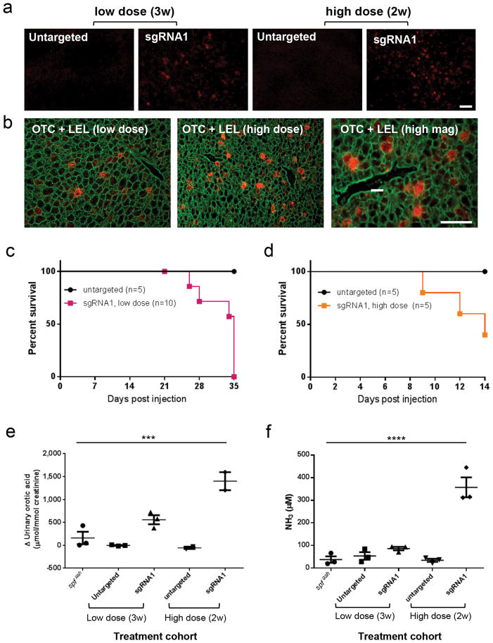
Many genetic liver diseases in newborns cause repeated, often lethal, metabolic crises. Gene therapy using nonintegrating viruses such as adeno-associated virus (AAV) is not optimal in this setting because the nonintegrating genome is lost as developing hepatocytes proliferate. We reasoned that newborn liver may be an ideal setting for AAV-mediated gene correction using CRISPR-Cas9. Here we intravenously infuse two AAVs, one expressing Cas9 and the other expressing a guide RNA and the donor DNA, into newborn mice with a partial deficiency in the urea cycle disorder enzyme, ornithine transcarbamylase (OTC). This resulted in reversion of the mutation in 10% (6.7-20.1%) of hepatocytes and increased survival in mice challenged with a high-protein diet, which exacerbates disease. Gene correction in adult OTC-deficient mice was lower and accompanied by larger deletions that ablated residual expression from the endogenous OTC gene, leading to diminished protein tolerance and lethal hyperammonemia on a chow diet.


Conflict of interest statement

Competing Financial Interests

J.M. Wilson is an advisor to REGENXBIO, Dimension Therapeutics, Solid Gene Therapy, and Alexion, and is a founder of, holds equity in, and has a sponsored research agreement with REGENXBIO and Dimension Therapeutics; in addition, he is a consultant to several biopharmaceutical companies and is an inventor on patents licensed to various biopharmaceutical companies.

Figures

Figure 1
Figure 1. In vivo gene correction of the OTC locus in the spfash mouse liver by AAV.CRISPR-SaCas9
(a) Schematic diagram of the mouse OTC locus showing the spfash mutation and three SaCas9 targets. spfash has a G-to-A mutation at the donor splice site at the end of exon 4 indicated in red on the top strand. The three selected SaCas9-targeted genomic sites (20 bp each) are in blue and underlined with the PAM sequences marked in green. The black line above exon 4 indicates the 1.8 kb OTC donor template. (b) Dual AAV vector system for liver-directed and SaCas9-mediated gene correction. The AAV8.sgRNA1.donor vector contains a 1.8-kb murine OTC donor template sequence as shown in (a) with the corresponding PAM sequence mutated and an AgeI site inserted. (c) Flowchart showing the key steps of AAV8.CRISPR-SaCas9-mediated gene correction in the neonatal OTC spfash model.
Figure 2
Figure 2. Efficient restoration of OTC expression in the liver of spfash mice treated at neonatal stage by AAV8.CRISPR-SaCas9-mediated gene correction
AAV8.SaCas9 (5×10¹⁰ GC/pup) and AAV8.sgRNA1.donor (5×10¹¹ GC/pup) were administrated to postnatal day 2 (p2) spfash pups via the temporal vein. spfash mice were sacrificed at 3 (n=5) or 8 weeks (n=8) after treatment. Untargeted spfash mice received AAV8.SaCas9 (5×10¹⁰ GC/pup) and AAV8.control.donor (5×10¹¹ GC/pup) at p2, and livers were harvested 8 weeks post treatment (n=6). Untreated WT (n=3) and spfash mice (n=3) were included as controls. (a) Immunofluorescence staining with antibodies against OTC on liver sections from spfash mice treated with the dual AAV vectors for CRISPR-SaCas9-mediated gene correction. Stained areas typically represent clusters of corrected hepatocytes. Untreated controls show livers from wild type, spfash heterozygous, and spfash hemizygous mice. Scale bar, 100μm. (b) Quantification of gene correction based on the percentage of area on liver sections expressing OTC by immunostaining as presented in panel a. (c) Random distribution of clusters of corrected hepatocytes along the portal-central axis shown by double immunostaining against OTC (red) and glutamine synthetase (GS, green), which is a marker of central veins (p, portal vein; c, central vein). Scale bars, 300 μm (upper panel) and 100 μm (lower panel). (d) Groups of corrected hepatocytes expressing OTC (red) shown by immunofluorescence on sections co-stained with fluorescein-labeled tomato lectin (Lycopersicon esculentum lectin, LEL; green) which outlines individual hepatocytes. Scale bar, 50μm. (e) OTC enzyme activity in the liver lysate of spfash mice at 3 and 8 weeks following dual vector treatment. (f) Quantification of OTC mRNA levels in the liver by RT-qPCR using primers spanning exons 4–5 to amplify wild-type OTC. Mean ± SEM are shown. * P < 0.05, ** P < 0.01, *** P < 0.001, Dunnett’s test.
Figure 3
Figure 3. Time course of SaCas9 expression following neonatal vector administration and functional improvement following high-protein diet challenge
(a) Immunostaining with antibodies against FLAG on liver sections from an untreated mouse or treated spfash mice at 1, 3, or 8 weeks following neonatal injection of the dual AAV vectors for CRISPR-SaCas9-mediated gene correction. AAV8.SaCas9 (5×10¹⁰ GC/pup) and AAV8.sgRNA1.donor (5×10¹¹ GC/pup) were administrated to p2 spfash pups via the temporal vein. Nuclear staining of FLAG-tagged SaCas9 were abundant at 1 week (n=5) but dramatically reduced at 3 weeks (n=6) and became scarce at 8 weeks (n=7) after vector injection. Scale bar, 100 μm. (b) Quantification of SaCas9 mRNA levels in liver by RT-qPCR. Mean ± SEM are shown. * P < 0.05, ** P < 0.01, *** P < 0.001, **** P < 0.0001, Dunnett’s test. (c) Quantification of SaCas9 vector genome in liver by qPCR. (d, e) Plasma ammonia levels and survival curves in control or dual AAV vector-treated spfash mice after a one-week course of high-protein diet. Seven weeks following neonatal treatment with the dual AAV vectors, mice were given high-protein diet for 7 days. (d) Plasma ammonia levels were measured 7 days after the high-protein diet. Plasma ammonia levels in WT mice (n=13) and AAV8.SaCas9 + AAV8.sgRNA1.donor-treated spfash mice (n=13) were significantly lower than untreated spfash mice (n=16) after a 7-day high-protein diet. Red squares indicate samples obtained from moribund untreated spfash mice 6 days after high-protein diet; red triangle indicates sample obtained from a moribund spfash mouse treated with untargeted vector (AAV8.control.donor with no sgRNA1, n=10) 5 days after high-protein diet. ** P< 0.01, **** P< 0.0001, Dunnett’s test. (e) Untreated spfash mice (n=20) or spfash mice treated with untargeted vectors (AAV8.control.donor, n=13) started to die 3 days after high-protein diet. All WT (n=13) and AAV8.SaCas9 + AAV8.sgRNA1.donor-treated mice (n=13) survived. * P< 0.05, Mantel-Cox test.
Figure 4
Figure 4. Gene targeting/correction in the liver of spfash mice treated as adults by AAV8.CRISPR-SaCas9 vectors
Adult spfash mice (8–10 weeks old) received an intravenous injection of AAV8.SaCas9 (1×10¹¹ GC) and AAV8.sgRNA1.donor (1×10¹² GC), or higher dose of AAV8.SaCas9 (1×10¹² GC) and AAV8.sgRNA1.donor (5×10¹² GC), or untargeted vectors at the equivalent doses. (a) Immunofluorescence staining with antibodies against OTC on liver sections collected at 3 (low-dose, n=3) or 2 weeks (high-dose, n=3) after injection. Stained cells typically showed as single corrected hepatocytes. Scale bar, 100μm. (b) Isolated corrected hepatocytes expressing OTC (red) shown by immunofluorescence on sections co-stained with fluorescein-labeled tomato lectin (LEL; green) which outlines individual hepatocytes. Scale bar, 50μm. (c) Survival curve of the low-dose cohorts: sgRNA1 (n=10) or untargeted vector at the same dose (n=5). (d) Survival curve of the high-dose cohorts: sgRNA1 (n=5) or untargeted vector at the same doses (n=5). The experiment was terminated at 14 days post vector injection. (e) Change of urine orotic acid levels in adult spfash mice after treatment with high-dose gene targeting vectors (n=3 for untreated spfash and low-dose groups; n=2 for high-dose groups) (f) Elevation of plasma NH3 levels in adult spfash mice after treatment with high-dose gene targeting vectors (n=3 for each group). Mean ± SEM are shown. *** P < 0.001, **** P < 0.0001, Dunnett’s test.

References

    1. Cunningham SC, Dane AP, Spinoulas A, Logan GJ, Alexander IE. Gene delivery to the juvenile mouse liver using AAV2/8 vectors. Mol Ther. 2008;16:1081–1088. - PubMed
    1. Wang L, Wang H, Bell P, McMenamin D, Wilson JM. Hepatic gene transfer in neonatal mice by adeno-associated virus serotype 8 vector. Hum Gene Ther. 2012;23:533–539. - PMC - PubMed
    1. Batshaw ML, Tuchman M, Summar M, Seminara J. A longitudinal study of urea cycle disorders. Mol Genet Metab. 2014;113:127–130. - PMC - PubMed
    1. Lichter-Konecki U, Caldovic L, Morizono H, Simpson K. Ornithine Transcarbamylase Deficiency (1993–2013) Pagon RA, Adam MP, Ardinger HH, et al., editors. GeneReviews®. ( http://www.ncbi.nlm.nih.gov/books/NBK154378/)
    1. Ah Mew N, Krivitzky L, McCarter R, Batshaw M, Tuchman M. Clinical outcomes of neonatal onset proximal versus distal urea cycle disorders do not differ. J Pediatr. 2013;162:324–329. e321. - PMC - PubMed

Publication types

MeSH terms

Substances