. 2016 Feb;34(2):204-9.
doi: 10.1038/nbt.3440. Epub 2016 Feb 1.

Cre-dependent selection yields AAV variants for widespread gene transfer to the adult brain

Affiliations

Cre-dependent selection yields AAV variants for widespread gene transfer to the adult brain

Benjamin E Deverman et al. Nat Biotechnol. 2016 Feb.

Abstract

Recombinant adeno-associated viruses (rAAVs) are commonly used vehicles for in vivo gene transfer. However, the tropism repertoire of naturally occurring AAVs is limited, prompting a search for novel AAV capsids with desired characteristics. Here we describe a capsid selection method, called Cre recombination-based AAV targeted evolution (CREATE), that enables the development of AAV capsids that more efficiently transduce defined Cre-expressing cell populations in vivo. We use CREATE to generate AAV variants that efficiently and widely transduce the adult mouse central nervous system (CNS) after intravenous injection. One variant, AAV-PHP.B, transfers genes throughout the CNS with an efficiency that is at least 40-fold greater than that of the current standard, AAV9 (refs. 14,15,16,17), and transduces the majority of astrocytes and neurons across multiple CNS regions. In vitro, it transduces human neurons and astrocytes more efficiently than does AAV9, demonstrating the potential of CREATE to produce customized AAV vectors for biomedical applications.


Figures

Figure 1
Figure 1. Cre-dependent recovery of AAV capsid sequences from transduced target cells
(a) An overview of the CREATE selection process. PCR is used to introduce diversity (full visual spectrum vertical band) into a capsid gene fragment (yellow). The fragment is cloned into the rAAV genome harboring the remaining capsid gene (gray) and is used to generate a library of virus variants. The library is injected into Cre transgenic animals and PCR is used to selectively recover capsid sequences from Cre+ cells. (b) The rAAV-Cap-in-cis-lox rAAV genome. Cre inverts the polyadenylation (pA) sequence flanked by the lox71 and lox66 sites. PCR primers (half arrows) are used to selectively amplify Cre-recombined sequences. (c) PCR products from Cre recombination-dependent (top) and -independent (bottom) amplification of capsid library sequences recovered from two Cre+ or Cre mice are shown. Schematics (bottom) show the PCR amplification strategies (see Supplementary Fig. 1 for details). (d) Schematic shows the AAV genes within the Rep-AAP AAV helper plasmid and the proteins encoded by the cap gene. Stop codons inserted in the cap gene eliminate VP1, VP2 and VP3 capsid protein expression. (e) DNase-resistant AAV vector genomes (vg) produced with the split AAV2/9 rep-AAP and rAAV-Cap-in-cis-lox genome (top) as compared to the vg produced with standard AAV2/9 rep-cap helper and rAAV-UBC-mCherry genome (middle) or with the AAV2/9 rep-AAP and rAAV-UBC-mCherry genome (bottom). N=3 independent trials per group; mean ± s.d.; **p<0.01, ***p<0.001; one-way ANOVA and Tukey multiple comparison test. (f) Cloning the 7-mer capsid library into the rAAV-ΔCap-in-cis vector. (g) The AAV9 surface model shows the location of the 7-mer inserted between AA588-589 (magenta). Sites encoded with the PCR-generated library fragment (AA450-592) are shown in yellow.
Figure 2
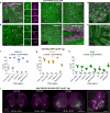
Figure 2. AAV-PHP.B mediates efficient gene delivery throughout the CNS after intravenous injection in adult mice
(a-f) ssAAV9:CAG-GFP or ssAAV-PHP.B:CAG-GFP, at 1×10¹² vg/mouse or 1×10¹¹ (a, right), was intravenously injected into adult mice. Images show GFP expression 3 weeks after injection. (a) Representative images of GFP IHC in the brains of mice given AAV9 (left) or AAV-PHP.B (middle and right). (b) Native GFP fluorescence in the cortex (left) or striatum (right) in 50 μm maximum intensity projection (MIP) confocal images. (c) GFP fluorescence in the PARS-cleared lumbar spinal cord. (d) GFP fluorescence in the retina (left: 20 μm MIP, transverse section; right: whole-mount MIP). (e,f) GFP fluorescence in 3D MIP images of PARS-cleared tissue from AAV-PHP.B transduced cortex and striatum (e) and indicated organs from mice transduced with AAV9 (top) or AAV-PHP.B (bottom) (f). Arrows highlight GFP+ nerves. Asterisks in the image of the pancreas highlight GFP+ islet cells. Major tick marks in 3D projections are 100 μm. (g) AAV biodistribution in the indicated brain regions (top) and organs (bottom) 25 days after intravenous injection of 1×10¹¹ vg into adult mice. N=3 for AAV-PHP.B and n=4 for AAV9; mean ± s.d.; **p<0.01, ***p<0.001, unpaired t tests corrected for multiple comparisons by the Holm-Sidak method. Scale bars: 1 mm (a, c (left)); 50 μm (b, c (right), d, e). Major tick marks in 3D projections in c, e, f are 100 μm.
Figure 3
Figure 3. AAV-PHP.B transduces multiple CNS cell types more efficiently than AAV9
(a-e) AAV-PHP.B transduces astrocytes, oligodendrocytes and neurons. Images show GFP expression 3 weeks after intravenous injection of 1×10¹¹ (a) or 1×10¹² (b-e) of ssAAV-PHP.B:CAG-GFP into adult mice. (a) MIP image of GFP (green) and GFAP (magenta) IHC in the hippocampus. (b) GFP and CC1 (magenta) IHC in the cortex. Numbered boxes highlight examples of double positive cells. Corresponding single-channel images are shown (right). Asterisks highlight cells without detectable GFP expression. (c) GFP and NeuN IHC in the indicated brain region. (d) MIP image of GFP fluorescence and TH IHC in the midbrain. (e) GFP and Calbindin (Calb) IHC in the cerebellum. (f-h) Quantification of the percentage of cells positive for NLS-GFP in the indicated brain region 3 weeks post injection. (i) AAV-PHP.B transduces ChAT+ spinal motor neurons. Images show native NLS-GFP fluorescence and IHC for ChAT in whole transverse spinal cord sections (left) or ventral horn MIP image (right). The percentage of ChAT+ neurons that expressed NLS-GFP in each spinal cord region is given ± the 95% confidence interval. For quantification, n=5 per group; mean ± s.d; All pairs of AAV9 vs AAV-PHP.B means were found to be different (***p<0.001) unpaired t tests corrected for multiple comparisons by the Holm-Sidak method. Scale bars: 20 μm (a,b,d), 50 μm (c,i, right), 200 μm (e) and 1 mm (i, left).
Figure 4
Figure 4. AAV-PHP.A exhibits efficient transduction of CNS astrocytes and reduced tropism for peripheral organs
(a-c, e) Images show GFP expression 3 weeks after intravenous injection of 3×10¹¹ of ssAAV9:CAG-GFP or ssAAV-PHP.A:CAG-GFP into adult mice. (a,b) Representative images of GFP IHC. (c) GFP IHC (green) and GFAP IHC (magenta) in the hippocampus. Numbered boxes highlight examples of GFP+/GFAP+ cells. Corresponding single-channel images are shown on the right. (d) 2.5×10¹¹ vg of ssAAV-PHP.A:CAG-NLS-GFP was injected intravenously into adult mice. Graphs show quantification of the percentage of Aldh1L1+ (blue) and NeuN+ cells (orange) positive for NLS-GFP. (e) Native GFP expression in the liver (green) and tissue autofluorescence (magenta). (f) AAV biodistribution in the brain (top) and peripheral organs (bottom) 25 days after intravenous injection of 1×10¹¹ vg into adult mice. (e, f) N=4 per group; mean ± s.d; *p<0.05, **p<0.01, ***p<0.001, unpaired t tests corrected for multiple comparisons by the Holm-Sidak method. Scale bars: 1 mm (a); 100 μm (b); 50 μm (c).

Similar articles

Cited by

References

    1. Kaplitt MG, et al. Safety and tolerability of gene therapy with an adeno-associated virus (AAV) borne GAD gene for Parkinson's disease: an open label, phase I trial. Lancet. 2007;369:2097–2105. doi:10.1016/S0140-6736(07)60982-9. - PubMed
    1. Wu Z, Asokan A, Samulski RJ. Adeno-associated virus serotypes: vector toolkit for human gene therapy. Molecular therapy : the journal of the American Society of Gene Therapy. 2006;14:316–327. doi:10.1016/j.ymthe.2006.05.009. - PubMed
    1. High KH, Nathwani A, Spencer T, Lillicrap D. Current status of haemophilia gene therapy. Haemophilia : the official journal of the World Federation of Hemophilia 20 Suppl. 2014;4:43–49. doi:10.1111/hae.12411. - PubMed
    1. Borel F, Kay MA, Mueller C. Recombinant AAV as a platform for translating the therapeutic potential of RNA interference. Molecular therapy : the journal of the American Society of Gene Therapy. 2014;22:692–701. doi:10.1038/mt.2013.285. - PMC - PubMed
    1. Ojala DS, Amara DP, Schaffer DV. Adeno-associated virus vectors and neurological gene therapy. Neuroscientist. 2015;21:84–98. doi:10.1177/1073858414521870. - PubMed

Publication types