

Enhanced Depth Imaging Optical Coherence Tomography in Uveitis: An Intravisit and Interobserver Reproducibility Study
- PMID: 26829594
- PMCID: PMC4811716
- DOI: 10.1016/j.ajo.2016.01.004
Enhanced Depth Imaging Optical Coherence Tomography in Uveitis: An Intravisit and Interobserver Reproducibility Study
Abstract
Purpose: To determine the intravisit and interobserver reproducibility of subfoveal choroidal thickness (SFCT) measurements in patients with noninfectious uveitis.
Design: Reliability analysis.
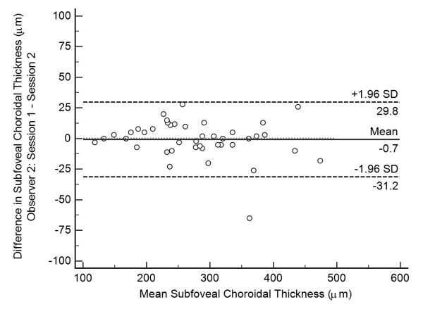
Methods: Two consecutive enhanced depth imaging optical coherence tomography (EDI-OCT) scans were obtained at a single clinic visit for 97 uveitic eyes from patients ≥16 years of age with noninfectious anterior (n = 10), intermediate (n = 11), posterior (n = 26), and panuveitis (n = 13) at the National Eye Institute. SFCT was manually measured by 2 ophthalmologists using manufacturer's software. Intravisit and interobserver reproducibility of SFCT measurements were assessed by using the Bland-Altman method to determine the estimate of bias (mean difference in SFCT measurements), 95% limits of agreement, and coefficients of repeatability. The reproducibility of these measurements was also compared between groups by anatomic location and clinical activity.
Results: Of 97 eyes, 65 (67.0%) were clinically quiet, 18 (18.6%) were minimally active, and 14 (14.4%) were active at the time the scans were obtained. Manual SFCT measurements were reproducible within 32.4 ± 3.8 μm between sessions for the same observer and 51.4 ± 8.5 μm between observers for the same session. Coefficients of repeatability did not differ significantly by anatomic location or disease activity.
Conclusions: Manual SFCT measurements obtained by EDI-OCT are reproducible in uveitis patients, with coefficients of repeatability that are nearly comparable to those published for normal eyes. This study provides guidance for using manual SFCT measurements in clinical practice, but further studies are still needed to determine their utility in clinical trials.
Published by Elsevier Inc.
Figures


Similar articles
-
Repeatability of manual subfoveal choroidal thickness measurements in healthy subjects using the technique of enhanced depth imaging optical coherence tomography.Invest Ophthalmol Vis Sci. 2011 Apr 8;52(5):2267-71. doi: 10.1167/iovs.10-6024. Print 2011 Apr. Invest Ophthalmol Vis Sci. 2011. PMID: 21087970
-
Reproducibility of subfoveal choroidal thickness measurements with enhanced depth imaging by spectral-domain optical coherence tomography.Invest Ophthalmol Vis Sci. 2013 Jan 9;54(1):230-3. doi: 10.1167/iovs.12-10351. Invest Ophthalmol Vis Sci. 2013. PMID: 23060144
-
Influence of choroidal thickness on subfoveal choroidal thickness measurement repeatability using enhanced depth imaging optical coherence tomography.Eye (Lond). 2014 Oct;28(10):1151-60. doi: 10.1038/eye.2014.197. Epub 2014 Sep 12. Eye (Lond). 2014. PMID: 25214002 Free PMC article.
-
Posterior Choroidal Stroma Reduces Accuracy of Automated Segmentation of Outer Choroidal Boundary in Swept Source Optical Coherence Tomography.Invest Ophthalmol Vis Sci. 2018 Sep 4;59(11):4404-4412. doi: 10.1167/iovs.18-24665. Invest Ophthalmol Vis Sci. 2018. PMID: 30193311
-
[Research progress of optical coherence tomography and optical coherence tomography angiography in noninfectious uveitis: a review].Zhonghua Yan Ke Za Zhi. 2023 Aug 11;59(8):677-681. doi: 10.3760/cma.j.cn112142-20220905-00433. Zhonghua Yan Ke Za Zhi. 2023. PMID: 37550977 Review. Chinese.
Cited by
-
Choroidal vascularity index as a biomarker of systemic inflammation in childhood Polyarteritis Nodosa and adenosine deaminase-2 deficiency.Pediatr Rheumatol Online J. 2020 Apr 3;18(1):29. doi: 10.1186/s12969-020-0417-3. Pediatr Rheumatol Online J. 2020. PMID: 32245490 Free PMC article.
-
Repeatability of Choroidal Thickness Measurements on Enhanced Depth Imaging Optical Coherence Tomography Using Different Posterior Boundaries.Am J Ophthalmol. 2016 Sep;169:104-112. doi: 10.1016/j.ajo.2016.06.023. Epub 2016 Jun 23. Am J Ophthalmol. 2016. PMID: 27345731 Free PMC article.
-
Choroidal thickness changes determined by EDI-OCT on acute anterior uveitis in patients with HLA-B27-positive ankylosing spondylitis.Int Ophthalmol. 2018 Feb;38(1):307-312. doi: 10.1007/s10792-017-0464-z. Epub 2017 Feb 14. Int Ophthalmol. 2018. PMID: 28197814
-
Optical coherence tomography: A guide to interpretation of common macular diseases.Indian J Ophthalmol. 2018 Jan;66(1):20-35. doi: 10.4103/ijo.IJO_902_17. Indian J Ophthalmol. 2018. PMID: 29283118 Free PMC article. Review.
-
Inter- and intraobserver repeatability and reproducibility of choroidal thickness measurements using two different methods.Int Ophthalmol. 2019 May;39(5):1061-1069. doi: 10.1007/s10792-018-0909-z. Epub 2018 Mar 31. Int Ophthalmol. 2019. PMID: 29605881
References
-
- Heidelberg Engineering. Spectral Domain Optical Coherence Tomography (SDOCT) [Accessed May 8, 2015]; Available at http://www.heidelbergengineering.com/us/products/spectralis-models/imagi...
-
- Spaide RF, Koizumi H, Pozzoni MC. Enhanced depth imaging spectral-domain optical coherence tomography. Am J Ophthalmol. 2008;146(4):496–500. - PubMed
-
- Bland JM, Altman DG. Statistical methods for assessing agreement between two methods of clinical measurement. Lancet. 1986;1(8476):307–310. - PubMed
-
- Rahman W, Chen FK, Yeoh J, Patel P, Tufail A, Da Cruz L. Repeatability of manual subfoveal choroidal thickness measurements in healthy subjects using the technique of enhanced depth imaging optical coherence tomography. Invest Ophthalmol Vis Sci. 2011;52(5):2267–2271. - PubMed
-
- Shao L, Xu L, Chen CX, et al. Reproducibility of subfoveal choroidal thickness measurements with enhanced depth imaging by spectral-domain optical coherence tomography. Invest Ophthalmol Vis Sci. 2013;54(1):230–233. - PubMed
Publication types
MeSH terms
Grants and funding
LinkOut - more resources
Full Text Sources
Other Literature Sources
