

Loss of Function of Intestinal IL-17 and IL-22 Producing Cells Contributes to Inflammation and Viral Persistence in SIV-Infected Rhesus Macaques
- PMID: 26829644
- PMCID: PMC4735119
- DOI: 10.1371/journal.ppat.1005412
Loss of Function of Intestinal IL-17 and IL-22 Producing Cells Contributes to Inflammation and Viral Persistence in SIV-Infected Rhesus Macaques
Abstract
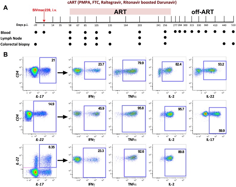
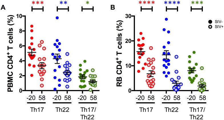
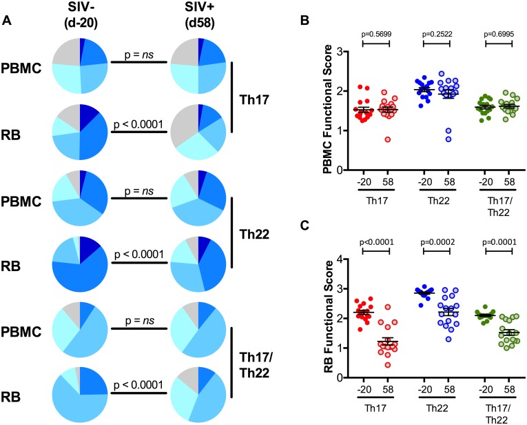
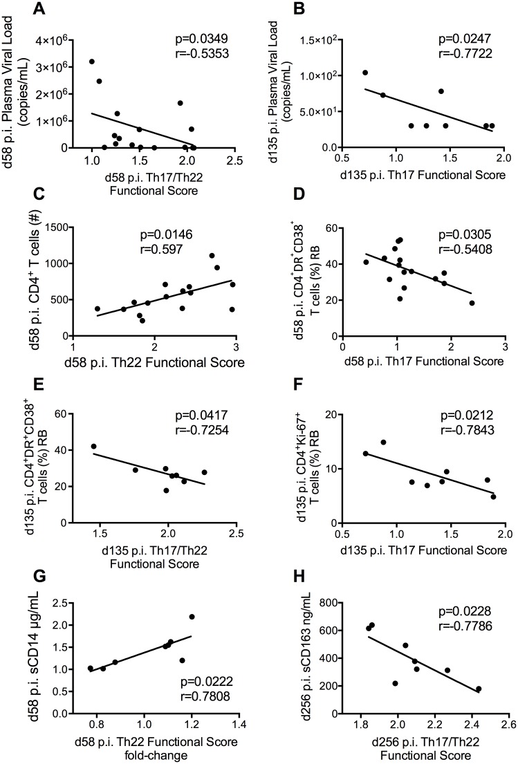
In HIV/SIV-infected humans and rhesus macaques (RMs), a severe depletion of intestinal CD4(+) T-cells producing interleukin IL-17 and IL-22 associates with loss of mucosal integrity and chronic immune activation. However, little is known about the function of IL-17 and IL-22 producing cells during lentiviral infections. Here, we longitudinally determined the levels and functions of IL-17, IL-22 and IL-17/IL-22 producing CD4(+) T-cells in blood, lymph node and colorectum of SIV-infected RMs, as well as how they recover during effective ART and are affected by ART interruption. Intestinal IL-17 and IL-22 producing CD4(+) T-cells are polyfunctional in SIV-uninfected RMs, with the large majority of cells producing four or five cytokines. SIV infection induced a severe dysfunction of colorectal IL-17, IL-22 and IL-17/IL-22 producing CD4(+) T-cells, the extent of which associated with the levels of immune activation (HLA-DR(+)CD38(+)), proliferation (Ki-67+) and CD4(+) T-cell counts before and during ART. Additionally, Th17 cell function during ART negatively correlated with residual plasma viremia and levels of sCD163, a soluble marker of inflammation and disease progression. Furthermore, IL-17 and IL-22 producing cell frequency and function at various pre, on, and off-ART experimental points associated with and predicted total SIV-DNA content in the colorectum and blood. While ART restored Th22 cell function to levels similar to pre-infection, it did not fully restore Th17 cell function, and all cell types were rapidly and severely affected--both quantitatively and qualitatively--after ART interruption. In conclusion, intestinal IL-17 producing cell function is severely impaired by SIV infection, not fully normalized despite effective ART, and strongly associates with inflammation as well as SIV persistence off and on ART. As such, strategies able to preserve and/or regenerate the functions of these CD4(+) T-cells central for mucosal immunity are critically needed in future HIV cure research.
Conflict of interest statement
The authors have declared that no competing interests exist.
Figures


Similar articles
-
Virologic and Immunologic Features of Simian Immunodeficiency Virus Control Post-ART Interruption in Rhesus Macaques.J Virol. 2020 Jul 1;94(14):e00338-20. doi: 10.1128/JVI.00338-20. Print 2020 Jul 1. J Virol. 2020. PMID: 32350073 Free PMC article.
-
Interleukin-21 combined with ART reduces inflammation and viral reservoir in SIV-infected macaques.J Clin Invest. 2015 Dec;125(12):4497-513. doi: 10.1172/JCI81400. Epub 2015 Nov 9. J Clin Invest. 2015. PMID: 26551680 Free PMC article.
-
Early Antiretroviral Therapy Prevents Viral Infection of Monocytes and Inflammation in Simian Immunodeficiency Virus-Infected Rhesus Macaques.J Virol. 2020 Oct 27;94(22):e01478-20. doi: 10.1128/JVI.01478-20. Print 2020 Oct 27. J Virol. 2020. PMID: 32907978 Free PMC article.
-
The Hitchhiker Guide to CD4+ T-Cell Depletion in Lentiviral Infection. A Critical Review of the Dynamics of the CD4+ T Cells in SIV and HIV Infection.Front Immunol. 2021 Jul 21;12:695674. doi: 10.3389/fimmu.2021.695674. eCollection 2021. Front Immunol. 2021. PMID: 34367156 Free PMC article.
-
Th17 cells and regulatory T cells in elite control over HIV and SIV.Curr Opin HIV AIDS. 2011 May;6(3):221-7. doi: 10.1097/COH.0b013e32834577b3. Curr Opin HIV AIDS. 2011. PMID: 21399494 Free PMC article. Review.
Cited by
-
Mucosal T Helper 17 and T Regulatory Cell Homeostasis Correlate with Acute Simian Immunodeficiency Virus Viremia and Responsiveness to Antiretroviral Therapy in Macaques.AIDS Res Hum Retroviruses. 2019 Mar;35(3):295-305. doi: 10.1089/AID.2018.0184. Epub 2019 Jan 2. AIDS Res Hum Retroviruses. 2019. PMID: 30398361 Free PMC article.
-
Inhibition of p38 MAPK in combination with ART reduces SIV-induced immune activation and provides additional protection from immune system deterioration.PLoS Pathog. 2018 Aug 30;14(8):e1007268. doi: 10.1371/journal.ppat.1007268. eCollection 2018 Aug. PLoS Pathog. 2018. PMID: 30161247 Free PMC article.
-
Virologic and Immunologic Features of Simian Immunodeficiency Virus Control Post-ART Interruption in Rhesus Macaques.J Virol. 2020 Jul 1;94(14):e00338-20. doi: 10.1128/JVI.00338-20. Print 2020 Jul 1. J Virol. 2020. PMID: 32350073 Free PMC article.
-
Human Th17 Cells Lack HIV-Inhibitory RNases and Are Highly Permissive to Productive HIV Infection.J Virol. 2016 Aug 12;90(17):7833-47. doi: 10.1128/JVI.02869-15. Print 2016 Sep 1. J Virol. 2016. PMID: 27334595 Free PMC article.
-
The Spontaneous Control of HIV Replication is Characterized by Decreased Pathological Changes in the Gut-associated Lymphoid Tissue.Curr HIV Res. 2018;16(5):338-344. doi: 10.2174/1570162X17666190130115113. Curr HIV Res. 2018. PMID: 30706820 Free PMC article.
References
-
- Veazey RS, DeMaria M, Chalifoux LV, Shvetz DE, Pauley DR, et al. (1998) Gastrointestinal tract as a major site of CD4+ T cell depletion and viral replication in SIV infection. Science 280: 427–431. - PubMed
Publication types
MeSH terms
Substances
Grants and funding
- NCRR P51RR000165/PHS HHS/United States
- P30 AI050409/AI/NIAID NIH HHS/United States
- P30AI50409/AI/NIAID NIH HHS/United States
- 5R24RR016988/RR/NCRR NIH HHS/United States
- P51 OD011132/OD/NIH HHS/United States
- P51 RR000165/RR/NCRR NIH HHS/United States
- R33AI104278/AI/NIAID NIH HHS/United States
- P51OD011132/OD/NIH HHS/United States
- R21AI116171/AI/NIAID NIH HHS/United States
- R33 AI104278/AI/NIAID NIH HHS/United States
- R01 AI084836/AI/NIAID NIH HHS/United States
- R01 AI110334/AI/NIAID NIH HHS/United States
- R24 RR016988/RR/NCRR NIH HHS/United States
- R33 AI116171/AI/NIAID NIH HHS/United States
- R01AI084836/AI/NIAID NIH HHS/United States
- R21 AI116171/AI/NIAID NIH HHS/United States
- R01AI110334/AI/NIAID NIH HHS/United States
LinkOut - more resources
Full Text Sources
Other Literature Sources
Research Materials
