Meta-Analysis
. 2016 Mar;48(3):273-82.
doi: 10.1038/ng.3500. Epub 2016 Feb 1.

MYB-QKI rearrangements in angiocentric glioma drive tumorigenicity through a tripartite mechanism

Pratiti Bandopadhayay  1   2   3   4 Lori A Ramkissoon  5 Payal Jain  6   7   8 Guillaume Bergthold  1   9 Jeremiah Wala  1   3   4 Rhamy Zeid  4   5 Steven E Schumacher  1   3 Laura Urbanski  1 Ryan O'Rourke  1   3 William J Gibson  1   3   4 Kristine Pelton  5 Shakti H Ramkissoon  5   10   11   12 Harry J Han  6   7 Yuankun Zhu  6   7 Namrata Choudhari  6   7 Amanda Silva  5   6   7 Katie Boucher  6   7 Rosemary E Henn  6   7 Yun Jee Kang  5 David Knoff  5 Brenton R Paolella  1   3   4 Adrianne Gladden-Young  13 Pascale Varlet  14 Melanie Pages  14 Peleg M Horowitz  1   15 Alexander Federation  4   5 Hayley Malkin  2 Adam A Tracy  3 Sara Seepo  3 Matthew Ducar  10   16 Paul Van Hummelen  16 Mariarita Santi  17   18 Anna Maria Buccoliero  19 Mirko Scagnet  20 Daniel C Bowers  21 Caterina Giannini  22 Stephanie Puget  23 Cynthia Hawkins  24 Uri Tabori  25 Almos Klekner  26 Laszlo Bognar  26 Peter C Burger  27 Charles Eberhart  27 Fausto J Rodriguez  27 D Ashley Hill  28   29   30 Sabine Mueller  31   32   33 Daphne A Haas-Kogan  32   34   35 Joanna J Phillips  32   36 Sandro Santagata  1   10   11   12 Charles D Stiles  1 James E Bradner  3   5   37 Nada Jabado  38   39   40 Alon Goren  13 Jacques Grill  9 Azra H Ligon  41 Liliana Goumnerova  2   42   43 Angela J Waanders  44   45   46   47 Phillip B Storm  6   7   45   47 Mark W Kieran  2   4 Keith L Ligon  3   5   10   11   12 Rameen Beroukhim  1   3   5   37   48 Adam C Resnick  6   7   45   49   47
Affiliations
Meta-Analysis

MYB-QKI rearrangements in angiocentric glioma drive tumorigenicity through a tripartite mechanism

Pratiti Bandopadhayay et al. Nat Genet. 2016 Mar.

Abstract

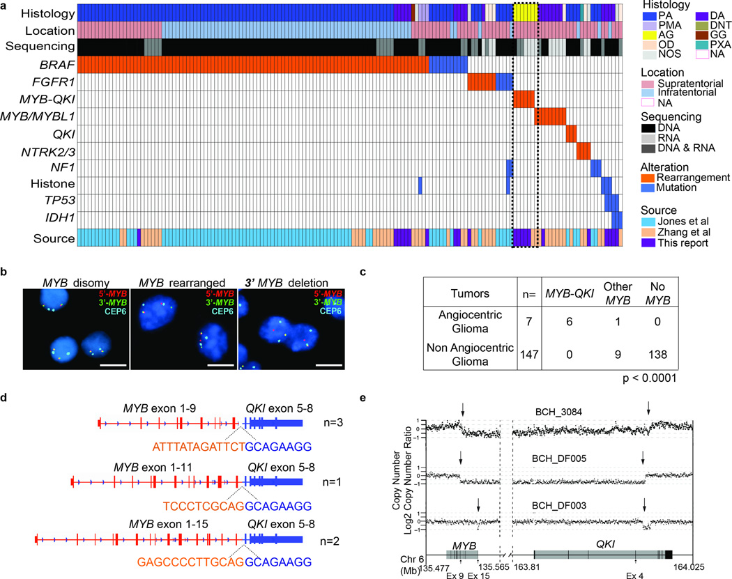
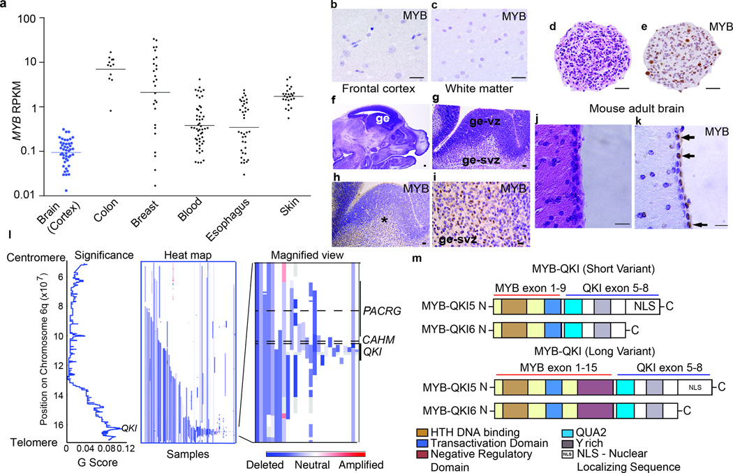
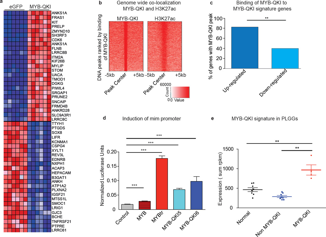
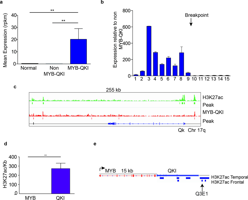
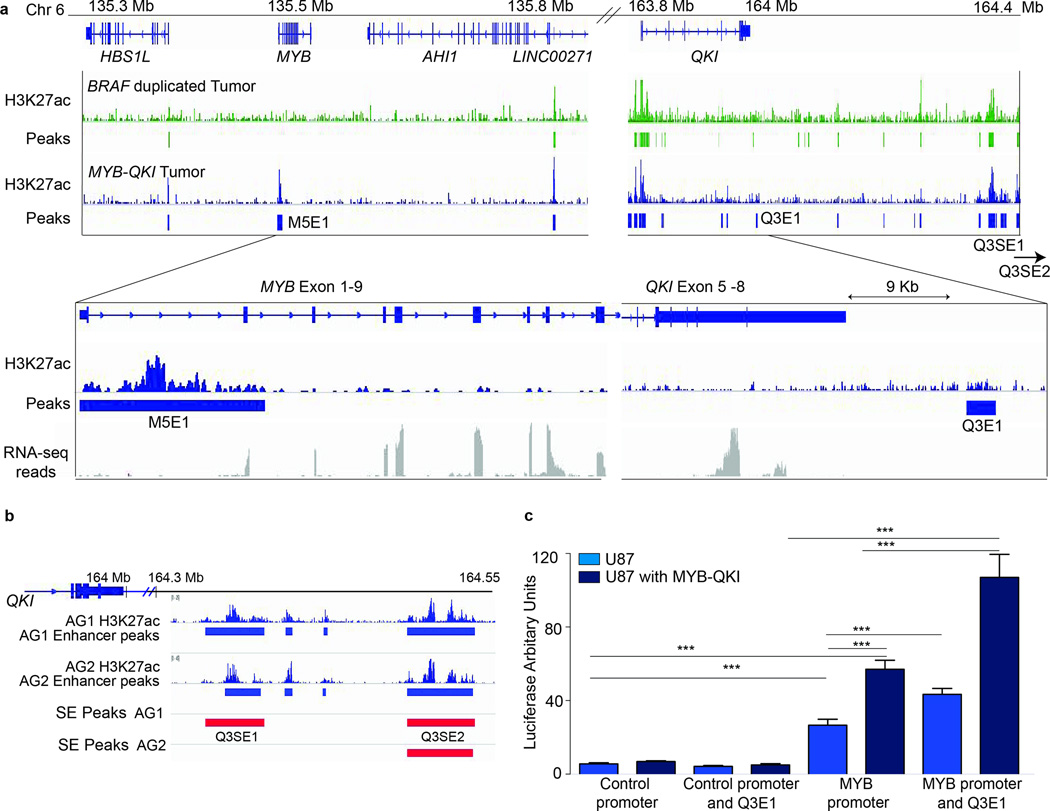
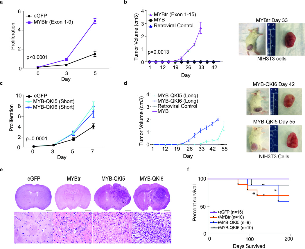
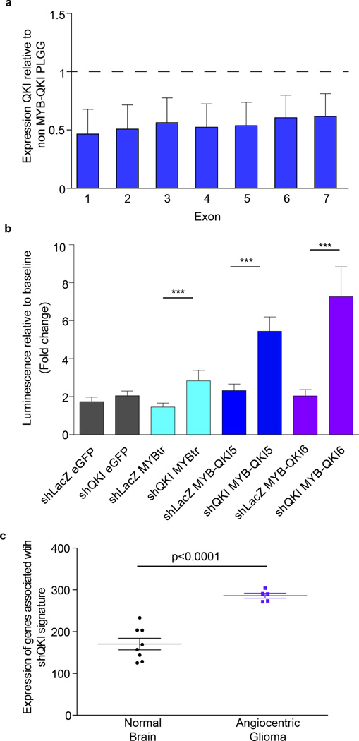
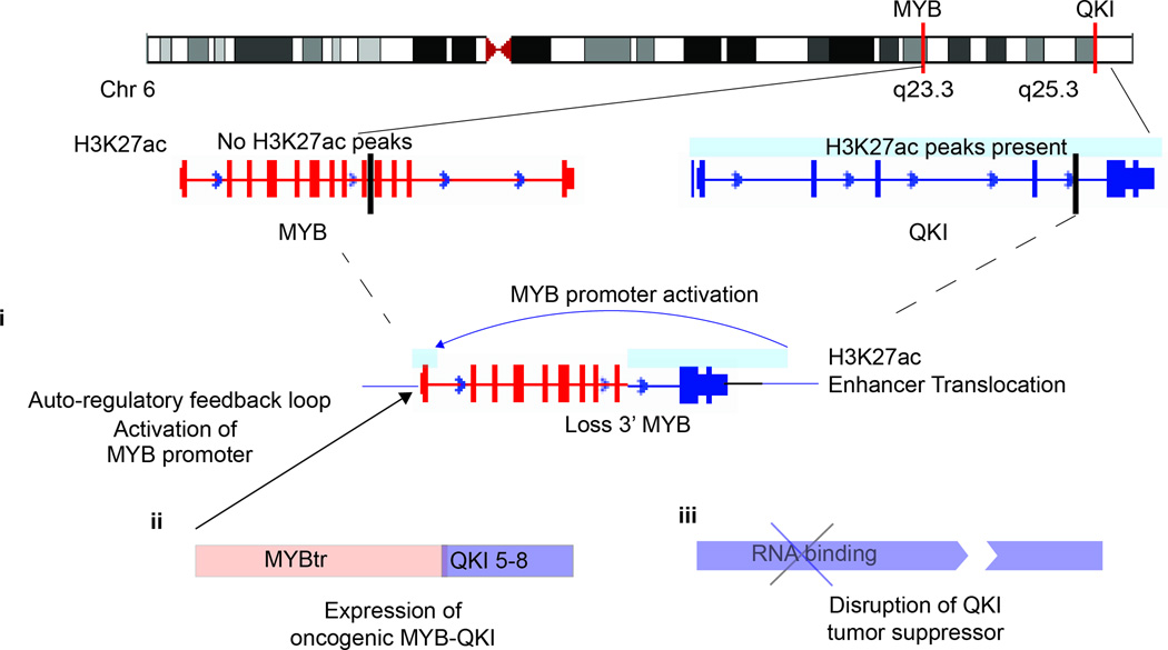
Angiocentric gliomas are pediatric low-grade gliomas (PLGGs) without known recurrent genetic drivers. We performed genomic analysis of new and published data from 249 PLGGs, including 19 angiocentric gliomas. We identified MYB-QKI fusions as a specific and single candidate driver event in angiocentric gliomas. In vitro and in vivo functional studies show that MYB-QKI rearrangements promote tumorigenesis through three mechanisms: MYB activation by truncation, enhancer translocation driving aberrant MYB-QKI expression and hemizygous loss of the tumor suppressor QKI. To our knowledge, this represents the first example of a single driver rearrangement simultaneously transforming cells via three genetic and epigenetic mechanisms in a tumor.


Conflict of interest statement

Conflict of interest statement: The authors declare no conflicts of interest.

Figures

Figure 1
Figure 1. Genomic analysis of 172 WGS and/or RNA-seq of PLGGs reveals a recurrent rearrangement involving MYB and QKI in Angiocentric Gliomas
a. Driver alterations were identified in 154 of 172 PLGGs profiled with WGS and/or RNA-seq. Histological subtypes include Pilocytic Astrocytoma (PA). Pilomyxoid Astrocytoma (PMA), Angiocentric Glioma (AG), Oligodendroglioma (OD), Diffuse Astrocytoma (DA), Dysembryoplastic Neuroepithelial Tumor (DNT), Ganglioglioma (GG), Pleomorphic Xanthoastrocytoma (PXA), and PLGG not otherwise specified (NOS). Tumors for which histology is unavailable are designated NA. b. FISH using probes flanking MYB reveal three patterns in PLGG: disomy, MYB rearrangement, or 3’ MYB deletion. Scale bars = 5 microns c. Frequency of MYB alterations or MYB-QKI rearrangements in Diffuse Astrocytoma and Angiocentric Glioma. p value represents enrichment of MYB-QKI rearrangements in Angiocentric Glioma. MYB-QKI alterations were identified with WGS alone (n=1), WGS and RNA-seq (n=2) or RNA-seq alone (n=3). d. Breakpoints observed in MYB and QKI in Angiocentric Gliomas. Sequence across the breakpoints as determined by RNA-seq is shown for each rearrangement. e. Copy-number profiles from WGS data of MYB and QKI in three Angiocentric Gliomas.
Figure 2
Figure 2. Alterations of MYB and QKI occur frequently in human cancers
a. MYB expression (mean ± SEM) in normal human colon (n=12), breast (n=27), whole blood (n=51), esophagus (n=38), skin (n=25), and brain cortex (n=47). b. MYB immunohistochemistry on human adult frontal cortex. Scale bar = 100 microns c. MYB immunohistochemistry on human adult white matter. Scale bar = 100 microns d. Hematoxylin and eosin (H&E) on human fetal neural stem cells generated from the ganglionic eminence at 22 weeks gestation. Scale bar = 100 microns e. MYB immunohistochemistry demonstrates positive staining in a subset of cells. Scale bar = 100 microns f. Sagittal section from embryonic 14.5 days post coitus (E14.5) mouse brain. Scale bar = 500 microns. g. H&E of E14.5 ganglionic eminence (ge) including ventricular (ge-vz) and subventricular (ge-svz) zones. Scale bar = 50 microns. h. MYB immunohistochemistry on the E14.5 ganglionic eminence. Scale bar = 50 microns. i. MYB immunohistochemistry demonstrates positive staining in subventricular zone (ge-svz) but not the ventricular zone (ge-vz). Scale bars = 50 microns. j. H&E from periventricular region of adult mouse brain. Scale bar = 100 microns. k. Immunohistochemistry for MYB demonstrates positive cells (arrows) in the ependymal/SVZ layer. Scale bars = 100 microns. l. (Left) Significance of deletions (x-axis) and (middle and right) heatmaps indicating copy-number profiles at 6q of individual adult Glioblastomas. m. Structure of the MYB-QKI fusion protein. TAD denotes transactivating domain. C-terminus of QKI includes QUA2 domains. MYB-QKI5 retains a nuclear localizing sequence (NLS). Two variants of MYB-QKI are depicted corresponding to the breakpoint of MYB.
Figure 3
Figure 3. MYB-QKI functions as a transcription factor, and its molecular effects are observed in Angiocentric Gliomas
a. MYB-QKI expression signature in mouse neural stem cells relative to cells expressing eGFP controls. b. Heatmap of H3K27ac and MYB-QKI levels at MYB-QKI regions. Each row is centered on MYB-QKI peaks. These regions are rank-ordered by MYB-QKI signal. Scaled intensities are in units of rpm/bp c. % of MYB-QKI signature genes with evidence of MYB-QKI ChIP-seq binding in up-regulated (n=25) and down-regulated (n=25) genes. ** depicts p<0.001 (paired t test). d. mim-1 reporter induction following transfection of MYBtr, MYB-QKI5, MYBQKI6 or full length MYB in 293T cells. Values shown represent mean of three independent measurements ± SEM. e. Expression of MYB-QKI signature in normal pediatric brain samples (n=8), PLGGs without MYB-QKI (n=8), or Angiocentric Gliomas with MYB-QKI (n=4). Values represent mean expression of signature in tumors ± SEM. Expression of signature within each tumor is the sum of rpkm of each gene in the signature.
Figure 4
Figure 4. Angiocentric gliomas exhibit aberrant expression of MYB-QKI due to H3K27ac-associated enhancer translocation and an autoregulatory feedback circuit in which MYB-QKI binds to the MYB promoter
a. MYB expression levels (in RPKM) of tumors with MYB-QKI rearrangement (n = 5) relative to normal brain (n = 10) or BRAF- or FGFR-driven PLGGs (n = 10). Values shown represent means ± s.e.m. **P < 0.05. b. Exon-specific expression of MYB in angiocentric gliomas that harbor MYB-QKI rearrangement (n = 3) relative to PLGGs that harbor BRAF alterations (n = 4). Values shown represent means ± s.e.m. c. Top track (green), H3K27ac binding within the Qk locus in mNSCs. Bottom track (red), MYB-QKI binding within the Qk locus in mNSCs. ChIP-seq binding peaks are shown. d. H3K27ac signal within the MYB and QKI loci in human frontal and temporal lobes (Encyclopedia of DNA Elements, ENCODE). Values shown depict the mean number of nucleotides that are associated with H3K27ac (per kb) in MYB and QKI across both locations ±s.e.m. (n = 1 ChIP-seq map for each location). **P < 0.05. e. Predicted H3K27ac-associated enhancer elements in MYB-QKI, with translocation of genomic enhancers from the 3’ region of QKI to within 15 kb of the 5’ end of MYB. The enhancer maps shown are derived from ENCODE data for normal human brain (frontal and temporal lobes). Q3E1 represents a H3K27ac-associated enhancer present in the ENCODE data from normal brain.
Figure 5
Figure 5. Human Angiocentric Gliomas exhibit H3K27ac enhancer translocation with an aberrant enhancer associated with the MYB promoter
a. H3K27ac enhancer peaks in proximity to MYB and QKI in a BRAF-duplicated Pilocytic Astrocytoma (top) and MYB-QKI Angiocentric Glioma (lower). Q3E1 is an enhancer associated with the 3’UTR of QKI. Two super-enhancer clusters (Q3SE1 and Q3SE2) are located within 500kb of QKI. Angiocentric Gliomas are associated with aberrant enhancer formation at the MYB promoter (M5E1), which is not detected in the BRAF driven pilocytic astrocytoma. The breakpoints for the MYB-QKI rearrangement are between exons 1–9 MYB and 5–8 QKI. Expression as determined by RNA-sequencing is depicted for the MYB-QKI Angiocentric Glioma. b. 3’ QKI associated super-enhancers (Q3SE1/2) presented in two Angiocentric Gliomas. c. MYB promoter activation following transfection of the MYB-luc construct in U87 cells and U87 cells over-expressing MYB-QKI with and without Q3E1 enhancer cloned into MYB-luc construct. Changes in luciferase activity of the MYB-luc reporter is shown as mean (± SEM) of three individual replicate experiments with n=5.
Figure 6
Figure 6. MYB-QKI fusion protein and truncated MYB are oncogenic
a. In vitro cell proliferation (number of cells relative to baseline) of mNSCs overexpressing eGFP or truncated MYBtrexons1–9. The mean values for five independent pools are depicted. Error bars, s.e.m. b. Tumor growth following flank injections of NIH3T3 cells overexpressing MYB, MYBtrexons1–15 or a vector control. The means of five measurements are depicted. Error bars, s.e.m. Representative images are shown for intracranial mNSC-MYB-QKI6 tumors. c. In vitro cell proliferation of mNSCs that overexpress MYB-QKI5 (short), MYB-QKI6 (short) or eGFP control. The means of five independent pools are depicted. Error bars, s.e.m. d. Tumor growth following flank injections of NIH3T3 cells overexpressing MYB, MYB-QKI5 (long), MYB-QKI6 (long) or vector control. The mean of five measurements is depicted. Error bars, s.e.m. Representative images are shown of intracranial mNSC–truncated MYB tumors. e. Hematoxylin and eosin analysis of severe combined immunodeficient (SCID) mouse brain after striatal injections with mNSCs expressing eGFP, truncated MYB, MYB-QKI5 or MYB-QKI6. Scale bars, 2 mm (top) and 50 ?m (bottom). f. Kaplan-Meier survival analysis of orthotopic SCID mice injected with mNSCs overexpressing truncated MYB, MYB-QKI5 or MYB-QKI6 that develop tumors with short latency in comparison to mice injected with mNSCs expressing eGFP, which never develop tumors (**P < 0.05).
Figure 7
Figure 7. MYB-QKI disrupts expression of QKI, a tumor suppressor gene
a. Exon specific expression of in Angiocentric Gliomas (n=5) relative to BRAF-driven PLGGs (n=5). Values represent mean ± SEM. RNA-sequencing data of Exon 8 of QKI revealed a high number of duplicate reads and thus is not shown. b. Cell proliferation of mouse neural stem cells expressing MYBtr, MYB-QKI5, MYB-QKI6 or eGFP control with suppression of wild-type Qk. Values represent mean of four independent experiments ± SEM. c. Expression of signature within each tumor is the sum of rpkm of each gene in the signature.
Figure 8
Figure 8. The MYB-QKI rearrangement contributes to oncogenesis through at least three mechanisms
The MYB-QKI rearrangement disrupts both MYB and QKI, resulting in hemizygous deletion of 3’MYB and 5’ QKI. This results in proximal translocation of H3K27ac enhancers on 3’QKI towards the MYB promoter, resulting in MYB promoter activation (i). The MYB-QKI fusion protein that is expressed is oncogenic, functions as a transcription factor, and exhibits the ability to bind to and activate the MYB promoter, resulting in an auto-regulatory feedback loop (ii). Hemizygous loss of QKI results in suppression of QKI expression, which functions as a tumor suppressor gene (iii).

References

References – Main Text

    1. Ramkissoon LA, et al. Genomic analysis of diffuse pediatric low-grade gliomas identifies recurrent oncogenic truncating rearrangements in the transcription factor MYBL1. Proc Natl Acad Sci U S A. 2013;110:8188–8193. - PMC - PubMed
    1. Zhang J, et al. Whole-genome sequencing identifies genetic alterations in pediatric low-grade gliomas. Nat. Genet. 2013;45:602–612. - PMC - PubMed
    1. Wang M, et al. Monomorphous angiocentric glioma: a distinctive epileptogenic neoplasm with features of infiltrating astrocytoma ependymoma. J. Neuropathol. Exp. Neurol. 2005;64:875–881. - PubMed
    1. Lellouch-Tubiana A, et al. Angiocentric neuroepithelial tumor (ANET): a new epilepsy-related clinicopathological entity with distinctive MRI. Brain Pathol. 2005;15:281–286. - PMC - PubMed
    1. Jones DTW, et al. Recurrent somatic alterations of FGFR1 NTRK2 in pilocytic astrocytoma. Nat. Genet. 2013;45:927–932. - PMC - PubMed

References – Online Methods

    1. Zhang J, et al. Whole-genome sequencing identifies genetic alterations in pediatric low-grade gliomas. Nat. Genet. 2013;45:602–612. - PMC - PubMed
    1. Jones DTW, et al. Recurrent somatic alterations of FGFR1 NTRK2 in pilocytic astrocytoma. Nat. Genet. 2013;45:927–932. - PMC - PubMed
    1. Li H, Durbin R. Fast and accurate short read alignment with Burrows-Wheeler transform. Bioinformatics. 2009;25:1754–1760. - PMC - PubMed
    1. McKenna A, et al. The Genome Analysis Toolkit: a MapReduce framework for analyzing next-generation DNA sequencing data. Genome Res. 2010;20:1297–1303. - PMC - PubMed
    1. Chiang DY, et al. High-resolution mapping of copy-number alterations with massively parallel sequencing. Nat. Methods. 2009;6:99–103. - PMC - PubMed

Publication types