Review
. 2016 Apr:37:115-22.
doi: 10.1016/j.sbi.2016.01.004. Epub 2016 Jan 30.

SPLINTS: small-molecule protein ligand interface stabilizers

Affiliations
Review

SPLINTS: small-molecule protein ligand interface stabilizers

Eric S Fischer et al. Curr Opin Struct Biol. 2016 Apr.

Abstract

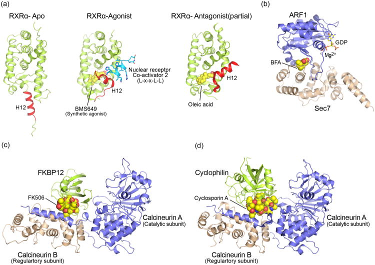
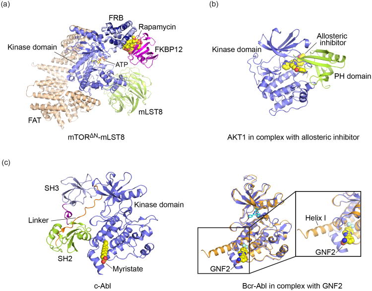
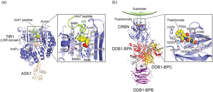
Regulatory protein-protein interactions are ubiquitous in biology, and small molecule protein-protein interaction inhibitors are an important focus in drug discovery. Remarkably little attention has been given to the opposite strategy-stabilization of protein-protein interactions, despite the fact that several well-known therapeutics act through this mechanism. From a structural perspective, we consider representative examples of small molecules that induce or stabilize the association of protein domains to inhibit, or alter, signaling for nuclear hormone, GTPase, kinase, phosphatase, and ubiquitin ligase pathways. These SPLINTS (small-molecule protein ligand interface stabilizers) drive interactions that are in some cases physiologically relevant, and in others entirely adventitious. The diverse structural mechanisms employed suggest approaches for a broader and systematic search for such compounds in drug discovery.


Figures

Figure 1
Figure 1
(a) Structural comparison among three different conformational state of nuclear receptor ligand binding domain. Activation domain is represented red helix in Apo (left, PDB ID: 1LBD), Agonist (middle, PDB ID: 1FBY) and Antagonist (Right, PDB ID: 2DKF). (b) Structure of Arf-Sec7 in complex with Brefeldin A (BFA). BFA is represented by space filling model (PDB ID: 1S9D). (c),(d) Structural comparison between FKBP-FK506-calcineurin (PDB ID:1TCO) and Cyclophilin-CyclosporinA-calcineurin(PDB ID:1M63), respectively.
Figure 2
Figure 2
Small molecules that inhibit kinases by inducing or stabilizing domain interactions. (a) Rapamycin recruits FKBP12 to mTOR, thereby restricting access of substrates to the kinase. The crystal structure of a partial mTOR complex revealed the structure and interactions of the mTOR FAT, kinase and FRB domains, and of the mLST8 subunit (shown in tan, blue and green, respectively, drawn from PDB ID 4JSP). Modeling of the interaction of rapamycin and FKBP12 with mTOR, based on superposition of the structure of an FRB:rapamycin:FKBP12 ternary complex (PDB ID 1FAP), showed how this induced protein interaction occupies a substrate binding surface on the FRB domain and constricts access to the kinase active site (shown with nucleotide substrate analog ATPγS bound). (b) Structure of human AKT in complex with allosteric inhibitor compound VIII (PDB ID: 3O96). The inhibitor stabilizes the interaction of the PH domain with the kinase domain, rendering it inactive due dismantling of key elements of the active site. (c) Allosteric Inhibition of Bcr-Abl by restoration of autoinhibitory interactions of the SH2 domain. In autoinhibited cAbl (PDB ID: 1OPK), insertion of the N-terminal myristoyl group (represented by myristate in this structure) into a pocket on the kinase domain promotes docking of the SH2 and SH3 domains onto the kinase domain. These interactions lock the kinase in an inactive conformation. In the oncogenic Bcr-Abl fusion protein, this myristoyl group is not present, but docking of the SH2 domain can be restored by small molecules such as GNF2, which mimic the action of myristate. Superposition of the Abl kinase domain determined in the presence of absence of GNF2 (right panel, blue and tan ribbons respectively) showed that the compound induces the “kinked” conformation of helix I required for the inhibitory SH2 domain interaction (drawn from PDB ID codes 2F4J and 3K5V).
Figure 3
Figure 3
(a) Structure of TIR1-ASK1 complex with auxin and IAA7 degron peptide (PDB ID: 2P1Q). A detailed view of the TIR1-auxin-IAA7 interface is shown in the inset, with key residues labeled. (b) Structure of DDB1-CRBN E3 ligase in complex with thalidomide (PDB ID: 4CI1). The predicted site of substrate recognition is indicated as schematically. Inset: Close up of the Thalidomide binding pocket in the CRBN-CTD with key residues depicted as sticks.

References

    1. Milroy LG, Grossmann TN, Hennig S, Brunsveld L, Ottmann C. Modulators of protein-protein interactions. Chem Rev. 2014;114:4695–4748. Comprehensive review of current methods and technologies available to the discovery of small molecule PPI modulators. Reviews broad examples of low molecular weight PPI stabilizers. - PubMed
    1. Fischer G, Rossmann M, Hyvonen M. Alternative modulation of protein-protein interactions by small molecules. Curr Opin Biotechnol. 2015;35:78–85. - PMC - PubMed
    1. Bier D, Thiel P, Briels J, Ottmann C. Stabilization of Protein-Protein Interactions in chemical biology and drug discovery. Prog Biophys Mol Biol. 2015;119:10–19. - PubMed
    1. Bourguet W, Germain P, Gronemeyer H. Nuclear receptor ligand-binding domains: three-dimensional structures, molecular interactions and pharmacological implications. Trends Pharmacol Sci. 2000;21:381–388. - PubMed
    1. Bourguet W, Ruff M, Chambon P, Gronemeyer H, Moras D. Crystal structure of the ligand-binding domain of the human nuclear receptor RXR-alpha. Nature. 1995;375:377–382. First structure of retinoid-X receptor (RXR) ligand-binding domain revealing its overall architecture and proposing the presence of a retionic acid binding pocket. - PubMed

Publication types