. 2016 Jun;11(2):279-93.
doi: 10.1007/s11481-016-9652-2. Epub 2016 Jan 30.

Neuropsychological, Neurovirological and Neuroimmune Aspects of Abnormal GABAergic Transmission in HIV Infection

Affiliations

Neuropsychological, Neurovirological and Neuroimmune Aspects of Abnormal GABAergic Transmission in HIV Infection

Tetyana Buzhdygan et al. J Neuroimmune Pharmacol. 2016 Jun.

Abstract

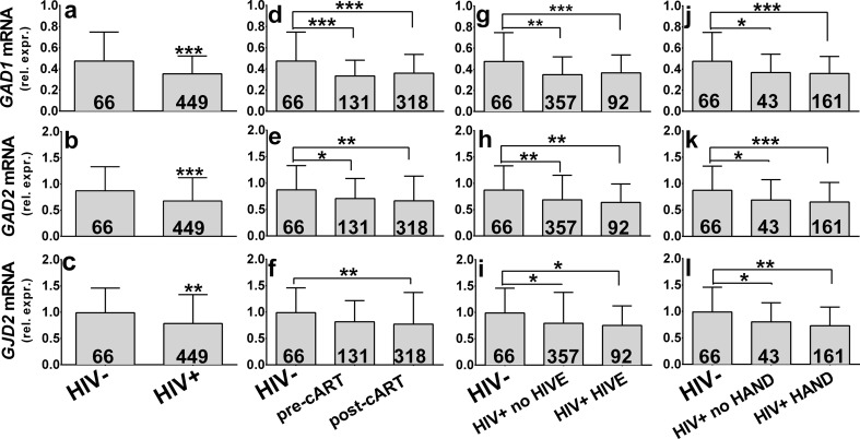
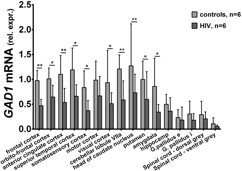
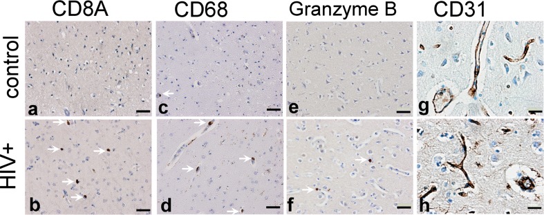
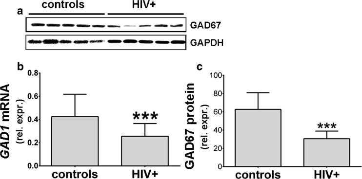
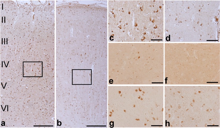
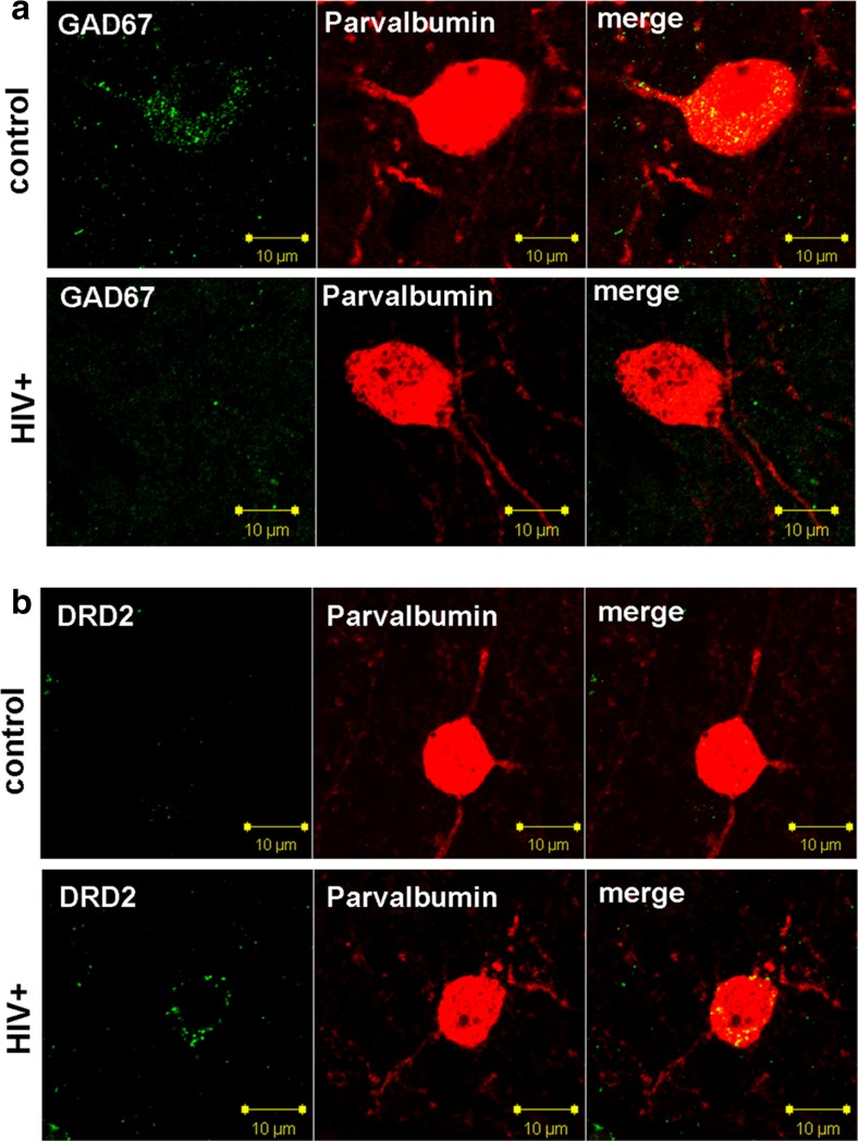
The prevalence of HIV-associated neurocognitive disorders (HAND) remains high in patients with effective suppression of virus replication by combination antiretroviral therapy (cART). Several neurotransmitter systems were reported to be abnormal in HIV-infected patients, including the inhibitory GABAergic system, which mediates fine-tuning of neuronal processing and plays an essential role in cognitive functioning. To elucidate the role of abnormal GABAergic transmission in HAND, the expression of GABAergic markers was measured in 449 human brain specimens from HIV-infected patients with and without HAND. Using real-time polymerase chain reaction, immunoblotting and immunohistochemistry we found that the GABAergic markers were significantly decreased in most sectors of cerebral neocortex, the neostriatum, and the cerebellum of HIV-infected subjects. Low GABAergic expression in frontal neocortex was correlated significantly with high expression of endothelial cell markers, dopamine receptor type 2 (DRD2L), and preproenkephalin (PENK) mRNAs, and with worse performance on tasks of verbal fluency. Significant associations were not found between low GABAergic mRNAs and HIV-1 RNA concentration in the brain, the history of cART, or HIV encephalitis. Pathological evidence of neurodegeneration of the affected GABAergic neurons was not present. We conclude that abnormally low expression of GABAergic markers is prevalent in HIV-1 infected patients. Interrelationships with other neurotransmitter systems including dopaminergic transmission and with endothelial cell markers lend added support to suggestions that synaptic plasticity and cerebrovascular anomalies are involved with HAND in virally suppressed patients.

Keywords: Autopsy; GABA; GABAergic; Glutamate decarboxylase; HIV associated neurocognitive disorders; HIV encephalitis.


Figures

Fig. 1
Fig. 1
Glutamate decarboxylase 1 (GAD1), glutamate decarboxylase 2 (GAD2), and connexin 36 (GJD2) mRNAs in the dorsolateral prefrontal cortex of 449 HIV infected and 66 HIV seronegative patients (Panels a - l). “Rel. exp.” on the ordinates denotes mRNA expression relative to GAPDH mRNA. The number of subjects in each group is given in the bars. Panels a - c. GABAergic mRNA expression was significantly lower in the HIV infected patients. Panels d – f. HIV-positive subjects were divided into subjects who died before and after the introduction of cART. mRNA expression was significantly lower in pre-cART and post-cART HIV patients as compared to the uninfected comparison group. There were no significant differences between pre-cART and post-cART groups. Panels g - i. HIV-positive subjects were divided according to neuropathological diagnosis of HIV-encephalitis (HIVE). All three mRNAs were significantly lower in patients without HIVE and with HIVE when compared to the uninfected comparison group. There were no significant differences between HIVE and no HIVE groups. Panels j - l. HIV-positive subjects were divided according to the diagnosis of HIV-associated neurocognitive disorders (HAND). Compared to the uninfected comparison group, all three mRNAs were significantly lower in patients without HAND and with HAND. There were no significant differences between HAND and no HAND groups. Mean ± standard deviation. *p < 0.05; **p < 0.01; ***p < 0.001
Fig. 2
Fig. 2
Glutamate decarboxylase 1 mRNA (GAD1) expression was measured in 16 different brain regions obtained from 6 HIV-infected patients and 6 uninfected patients. Using two-way ANOVA, only variation in GAD1 mRNA concentrations between brain regions was significant (F (16, 218) = 7.17, p < 10−4). GAD1 was lower in the HIV infected brain specimens in most regions including neocortex, cerebellum and neostriatum (caudate nucleus and putamen). Uncorrected Fisher’s LSD tests were significant for dorsolateral prefrontal (- 51.7 %, p = 0.010), anterior cingulate (- 51.2 %, p = 0.009), superior temporal (- 44.5 %, p = 0.013), somatosensory (- 55.6 %, p = 0.042), and visual cortices (- 45.4 %, p = 0.038), cerebellar lobule VIIa (- 51.5 %, p = 0.003), head of caudate nucleus (- 42.5 %, p = 0.009), putamen (- 40.1 %, p = 0.049), and amygdala (- 60 %, p = 0.012). Mean decrease of GAD1 mRNA expression in the orbitofrontal cortex was - 36.2 % and almost reached significance (p = 0.0728). Spinal cord, paleostriatum, and hippocampus also had slightly lower GAD1 in the HIV infected brain specimens, but those differences were not significant statistically. Relative expression of GAD1 mRNA was normalized to GAPDH mRNA. Mean ± standard deviation. *p < 0.05; **p < 0.01; ***p < 0.001
Fig. 3
Fig. 3
Neuroimmune and endothelial cells were immunostained in the dorsolateral prefrontal cortex of the HIV-seronegative control (panels a, c, e, g) and HIV-infected subjects (b, d, f, h). CD8+ cytotoxic T cells (arrows, a vs b) and CD68+ macrophages (arrows, c vs d) were more numerous in specimens from HIV-infected subjects with low GAD1 expression, as suggested by concentrations of the corresponding mRNAs (see Table 2). Granzyme B-containing inflammatory cells were more numerous in specimens from HIV- infected subjects with low GAD1 expression (arrows, e vs f). Staining for the endothelial cell marker CD31 produced more intense marking of blood vessel walls (arrows) in HIV- infected subjects with low GAD1 (g vs h). Bars represent 50 um in af, and 20 um in g and h
Fig. 4
Fig. 4
Glutamate decarboxylase 67 (GAD67) protein and mRNA expression in the dorsolateral prefrontal cortex. GAD67 immunoblotting was done on 36 HIV-infected subjects and 12 seronegative controls. GAPDH blots were done for loading controls. Equal amounts of protein were loaded to each well. GAD67 band intensities were significantly lower in 36 HIV+ subjects compared to 12 HIV- subjects (-51.2 %, p = 0.0001, panel c). Band intensities for ten representative subjects are illustrated in panel a. GAD1 mRNA expression is shown for comparison (b), mean GAD1 mRNA decrease is -39,8 %, (p = 0.001). GAD67 is expressed as relative to the GAPDH band intensity. GAD1 is expressed as relative to GAPDH mRNA. Mean ± standard deviation is shown. P values were obtained using the Student’s t test. *p < 0.05; **p < 0.01; ***p < 0.001
Fig. 5
Fig. 5
Glutamate decarboxylase 67 (GAD67) immunoreactivity in brain tissue from an HIV infected patient (b, d, f, h) and a seronegative control (a, c, e, g). Panels a and b illustrate dorsolateral prefrontal cortex (DLPFC) with neocortical laminae labeled I through VI at left. The specimen from the HIV infected subject (b) contains less intense staining in all of the laminae compared to the seronegative patient (a). Boxed areas in a and b are magnified in c and d and illustrate less intense immunostaining in pyramidal cell cytoplasm, in pear-shaped interneurons and in neuritic processes. In the neostriatum (caudate nucleus), decreased GAD67 immunostaining in the HIV infected subject was not as clear-cut but still was present (e vs f). The neurons in the globus pallidus also were stained less intensely in the HIV infected subject. Note that the number of stained neurons in the HIV infected subject is similar to the seronegative control (g vs h). Scale bars are 200 um in a and b, and 100 um in cf
Fig. 6
Fig. 6
The six panels in a illustrate that loss of GAD67 staining in HIV infected patients does not correspond to the dropping out of interneurons. Immunofluorescence for interneuronal markers GAD67 and parvalbumin (PV) show that the cortical interneuron from the HIV-infected subject contains less intense GAD67 staining compared to the seronegative control. PV immunoreactivity of interneurons and neuritic processes is of equivalent intensity in these two specimens. The six panels in b illustrate that HIV-infected patients with HAND had high expression of dopamine receptor 2 (DRD2) in cortical interneurons, when interneuron from a seronegative patient lacks DRD2 staining. PV immunoreactivity of interneurons and neuritic processes is of equivalent intensity in these two specimens. Scale bars are 10 um

Similar articles

Cited by

References

    1. Akbarian S, Kim JJ, Potkin SG, Hagman JO, Tafazzoli A, Bunney WE, Jones EG. Gene expression for glutamic acid decarboxylase is reduced without loss of neurons in prefrontal cortex of schizophrenics. Arch Gen Psychiatry. 1995;52:258–266. doi: 10.1001/archpsyc.1995.03950160008002. - DOI - PubMed
    1. Ances BM, Vaida F, Cherner M, Yeh MJ, Liang CL, Gardner C, Grant I, Ellis RJ, Buxton RB, HIV Neurobehavioral Research Center (HNRC) Group HIV and chronic methamphetamine dependence affect cerebral blood flow. J Neuroimmune Pharmacol. 2011;6:409–419. doi: 10.1007/s11481-011-9270-y. - DOI - PMC - PubMed
    1. Anneken JH, Cunningham JI, Collins SA, Yamamoto BK, Gudelsky GA. MDMA increases glutamate release and reduces parvalbumin-positive GABAergic cells in the dorsal hippocampus of the rat: role of cyclooxygenase. J Neuroimmune Pharmacol. 2013;8:58–65. doi: 10.1007/s11481-012-9420-x. - DOI - PMC - PubMed
    1. Baldo JV, Schwartz S, Wilkins D, Dronkers NF. Role of frontal versus temporal cortex in verbal fluency as revealed by voxel-based lesion symptom mapping. J Int Neuropsychol Soc. 2006;12:896–900. doi: 10.1017/S1355617706061078. - DOI - PubMed
    1. Ballabh P, Braun A, Nedergaard M. The blood-brain barrier: an overview: structure, regulation, and clinical implications. Neurobiol Dis. 2004;16:1–13. doi: 10.1016/j.nbd.2003.12.016. - DOI - PubMed

Publication types

MeSH terms

Substances