. 2016 Dec;3(1):3.
doi: 10.1186/s40348-016-0031-0. Epub 2016 Jan 29.

Towards understanding microvillus inclusion disease

Affiliations

Towards understanding microvillus inclusion disease

Georg F Vogel et al. Mol Cell Pediatr. 2016 Dec.

Abstract

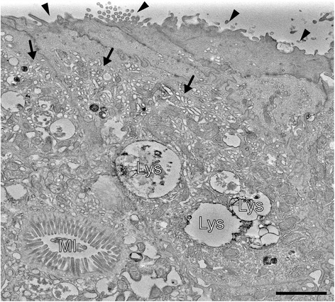
Microvillus inclusion disease (MVID) is characterised by onset of intractable life-threatening watery diarrhoea during infancy. Transmission electron microscopy demonstrates shortening or absence of apical microvilli, pathognomonic microvillus inclusions in mature enterocytes and subapical accumulation of periodic acid-Schiff-positive granules or vesicles confirming diagnosis. Mutations in MYO5B have been found to cause MVID. In two patients with MVID, whole-exome sequencing of DNA revealed homozygous truncating mutations in STX3. Mutations in these genes disrupt trafficking between apical cargo vesicles and the apical plasma membrane. Thus, disturbed delivery of certain brush border membrane proteins is a common defect in MVID.

Keywords: Enteropathy; MVID; MYO5B; Microvillus inclusion disease; STX3.


Figures

Fig. 1
Fig. 1
Electron micrograph of patient’s duodenal enterocytes depicting the three ultrastructural hallmarks of MVID (homozygous c.1323–2A > G splice-site mutation in MYO5B). Black arrow heads mark the shortening or loss of apical microvilli. Subapical accumulations of tubulo-/vesicular structures (secretory granules) are marked by black arrows. MI intracellular microvillus inclusion, Lys lysosomes. Scale bar = 2 μm

References

    1. Davidson GP, Cutz E, Hamilton JR, Gall DG. Familial enteropathy: a syndrome of protracted diarrhea from birth, failure to thrive, and hypoplastic villus atrophy. Gastroenterology. 1978;75(5):783–790. - PubMed
    1. Phillips AD, Jenkins P, Raafat F, Walker-Smith JA. Congenital microvillous atrophy: specific diagnostic features. Arch Dis Child. 1985;60(2):135–140. doi: 10.1136/adc.60.2.135. - DOI - PMC - PubMed
    1. Cutz E, Rhoads JM, Drumm B, Sherman PM, Durie PR, Forstner GG. Microvillus inclusion disease: an inherited defect of brush-border assembly and differentiation. N Engl J Med. 1989;320(10):646–651. doi: 10.1056/NEJM198903093201006. - DOI - PubMed
    1. Phillips AD, Schmitz J. Familial microvillous atrophy: a clinicopathological survey of 23 cases. J Pediatr Gastroenterol Nutr. 1992;14(4):380–396. doi: 10.1097/00005176-199205000-00003. - DOI - PubMed
    1. Phillips AD, Szafranski M, Man LY, Wall WJ. Periodic acid-Schiff staining abnormality in microvillous atrophy: photometric and ultrastructural studies. J Pediatr Gastroenterol Nutr. 2000;30(1):34–42. doi: 10.1097/00005176-200001000-00015. - DOI - PubMed