. 2016 Dec;21(12):1790-1798.
doi: 10.1038/mp.2015.228. Epub 2016 Feb 2.

Co-expression of truncated and full-length tau induces severe neurotoxicity

Affiliations

Co-expression of truncated and full-length tau induces severe neurotoxicity

S Ozcelik et al. Mol Psychiatry. 2016 Dec.

Abstract

Abundant tau inclusions are a defining hallmark of several human neurodegenerative diseases, including Alzheimer's disease. Protein fragmentation is a widely observed event in neurodegenerative proteinopathies. The relevance of tau fragmentation for the neurodegenerative process in tauopathies has yet remained unclear. Here we found that co-expression of truncated and full-length human tau in mice provoked the formation of soluble high-molecular-weight tau, the failure of axonal transport, clumping of mitochondria, disruption of the Golgi apparatus and missorting of synaptic proteins. This was associated with extensive nerve cell dysfunction and severe paralysis by the age of 3 weeks. When the expression of truncated tau was halted, most mice recovered behaviorally and functionally. In contrast, co-expression of full-length tau isoforms did not result in paralysis. Truncated tau thus induces extensive but reversible neurotoxicity in the presence of full-length tau through the formation of nonfilamentous high-molecular-weight tau aggregates, in the absence of tau filaments. Targeting tau fragmentation may provide a novel approach for the treatment of human tauopathies.


Figures
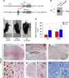
Figure 1
Figure 1
TAU62 mice express Δtau, develop a mild motor phenotype, memory deficits and pretangle pathology. (a) Expression constructs. In the presence of doxycycline, 3R tau151–421 (Δtau) is expressed. In the absence of doxycycline, tTS (tetracycline-controlled transcriptional silencer) binds to TRE (tetracycline-responsive element), preventing the expression of Δtau. (b) Western blot of brain using anti-tau antibody C3 of a 1-month-old TAU62 mouse under doxycycline (Dox on) and 3 days after doxycycline withdrawal (Dox on-off). (c) Tail suspension test on young TAU62 mouse (1 month), aged B6 mouse (18 months) and aged TAU62 mouse (18 months). (d) Object recognition test. Object exploration time of adult TAU62 mice (aged 6 months) and their C57Bl6 littermates. (e–m) Histology of TAU62 mice aged 12 months (e–g) and 18 months (h–m). Immunohistochemistry with C3 of somatomotor cortex and orbital area (e), hippocampus (f), brainstem with tegmental reticular nucleus (g) and spinal cord (k). Immunohistochemistry of brainstem with AT8 (h) and AT100 (i); immunohistochemistry of spinal cord with AT8 (l). Holmes–Luxol (HL) staining shows the presence of spheroids in spinal cord (arrow; inset) (m). The scale bar in (m) corresponds to 60 μm in (h, l and m), 80 μm in (i), 200 μm in (e) and 400 μm in (f, g and k). *P<0.05.
Figure 2
Figure 2
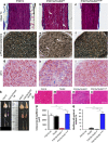
Co-expression of 4R P301S tau and Δtau (P301SxTAU62 mice) causes nerve cell dysfunction that is reversible upon cessation of Δtau expression. Paralysis is associated with the presence of soluble high-molecular-weight tau in the absence of sarkosyl-insoluble tau and tau filaments. Paralyzed mice exhibit axonal accumulations of neurofilaments and mitochondria. (a) Heterozygous P301S mouse (aged 3 weeks); paralyzed (aged 3 weeks) and recovered (3 weeks after cessation of Δtau expression) P301SxTAU62 mice (see also Supplementary Videos S1 and S3). (b) Recovery of motor function was assessed by a grid test of P301SxTAU62 mice following the removal of doxycycline at 21 days of age (blue lines). Motor function of heterozygous P301S tau littermates (green line, n=8). (c) Immunohistochemistry with AT8 of the tegmental reticular nucleus of the brainstem of paralyzed (‘on') and recovered (‘on-off') P301SxTAU62 mice; immunohistochemistry with AT100 (d), Gallyas–Braak silver (e) and Thioflavin S staining (f) of the reticular nucleus of paralyzed mice. (g) Western blot with human-specific anti-tau antibody HT7 of brainstem tissue from nontransgenic (B6), TAU62, P301S and P301SxTAU62 mice. (h and i) Holmes–Luxol (HL) staining of spinal cord of paralyzed 3-week-old P301SxTAU62 mice. The arrow in (i) points to a spheroid; (k) immunohistochemistry of paralyzed (‘on') and recovered (‘on-off') mice using antibodies against the 200 kDa subunit of neurofilaments (NF200). The scale bar in (c) corresponds to 26 μm in (h), 40 μm in (f and i), 80 μm in (k), 100 μm in (e) and 200 μm in (c and d). (l–n) Electron microscopy of the spinal cord of paralyzed mice. Only a few isolated microtubules are present in axons (m, arrow). Fragmented Golgi material is seen in nerve cell bodies (n, arrows). The scale bar in (l) corresponds to 5 nm in (l), 220 nm in (m) and 1.4 nm in (n).
Figure 3
Figure 3
Co-expression of 4R P301S tau and Δtau (P301SxTAU62 mice) causes neuropathy and neurogenic muscle atrophy that are reversible upon cessation of Δtau expression. (a–i) Sciatic nerves stained using Masson's trichrom stain (a–c), para-phenylenediamine (d–f) and 2f11 immunohistochemistry (g–i). The scale bar in (i) corresponds to 50 μm in (a–c), 32 μm in (d-f) and 25 μm in (g–i). (k) Macroscopic view of hindlimb muscles. From the top: M. gastrocnemius and M. soleus; M. tibialis anterior; M. extensor digitorum longus. (l–o) M. gastrocnemius stained with hematoxylin and eosin (HE). The scale bar in (o) corresponds to 100 μm (for l–o). Quantification of myofiber area (p) and myofibers with internalized nucleus (q). P301S: heterozygous mice transgenic for human mutant P301S tau, aged 3 weeks; TAU62: heterozygous mice expressing 3R tau151–421, aged 3 weeks; P301SxTAU62on: paralyzed mice, aged 3 weeks; P301SxTAU62on-off: recovered mice, 6 weeks after cessation of the expression of Δtau. ***P<0.001.
Figure 4
Figure 4
Co-expression of 4R wild-type tau and Δtau (ALZ17xTAU62) causes paralysis and neuropathy that are reversible upon cessation of Δtau expression. (a) Paralyzed (aged 3 weeks) and recovered (3 weeks after cessation of Δtau expression) ALZ17xTAU62 mice (see also Supplementary Videos S4 and S5). (b) Recovery of motor function as assessed by a grid test of ALZ17xTAU62 mice following the removal of doxycycline between 16 and 20 days of age (blue lines). Motor function of heterozygous ALZ17 littermates (green line) (n=6). (c) Western blot with HT7 of brainstem tissue from nontransgenic (B6), TAU62, ALZ17 and ALZ17xTAU62 mice. Actin staining was used as the loading control. (d–l) Histological analysis of paralyzed (‘on', d, f, g, h and k) and recovered (‘on-off', e, i and l) ALZ17xTAU62 mice using anti-tau antibody AT8 (d and e), anti-neurofilament antibody 2f11 (f), Masson's trichrome (h and i), Holmes–Luxol (HL) (g) and hematoxylin–eosin (HE) (k and l). The scale bar in (l) corresponds to 50 μm in (h, i, g, k and l), 100 μm in (f) and 200 μm in (d and e).
Figure 5
Figure 5
Co-expression of 4R mutant or wild-type tau and 3R wild-type (P301SxALZ31 or ALZ17xALZ31 mice) does not cause paralysis and results in pretangle pathology. (a and b) Unimpaired P301SxALZ31 and ALZ17xALZ31 mice aged 3 weeks (see also Supplementary Videos S7 and S8). (c) Western blot with HT7 of brainstem tissue from paralyzed ALZ31xTAU62 mice aged 3 weeks, unimpaired P301SxALZ31 aged 3 weeks and unimpaired ALZ17xALZ31 mice aged 4 months. Actin staining was used as the loading control. (d–l) Immunohistochemistry of 9-month-old P301SxALZ31 mouse and 4-month-old ALZ17xALZ31 mouse with AT8 ((d and h) cortex, (e and i) brainstem, (f and k) hippocampus and (g and l) spinal cord). The scale bar in (l) corresponds to 200 μm in (d, e, h and i), 500 μm in (f and k) and 50 μm in (g and l).

References

    1. Spillantini MG, Goedert M. Tau pathology and neurodegeneration. Lancet Neurol 2013; 12: 609–622. - PubMed
    1. Spires-Jones TL, Hyman BT. The intersection of amyloid beta and tau at synapses in Alzheimer's disease. Neuron 2014; 82: 756–771. - PMC - PubMed
    1. Frost B, Hemberg M, Lewis J, Feany MB. Tau promotes neurodegeneration through global chromatin relaxation. Nat Neurosci 2014; 17: 357–366. - PMC - PubMed
    1. Rosenmann H. Asparagine endopeptidase cleaves tau and promotes neurodegeneration. Nat Med 2014; 20: 1236–1238. - PubMed
    1. Eisenberg D, Jucker M. The amyloid state of proteins in human diseases. Cell 2012; 148: 1188–1203. - PMC - PubMed

Publication types