. 2016 Mar 31;36(8):1222-36.
doi: 10.1128/MCB.01035-15. Print 2016 Apr.

Arrested Hematopoiesis and Vascular Relaxation Defects in Mice with a Mutation in Dhfr

Affiliations

Arrested Hematopoiesis and Vascular Relaxation Defects in Mice with a Mutation in Dhfr

Julie A I Thoms et al. Mol Cell Biol. .

Abstract

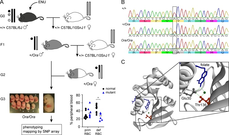
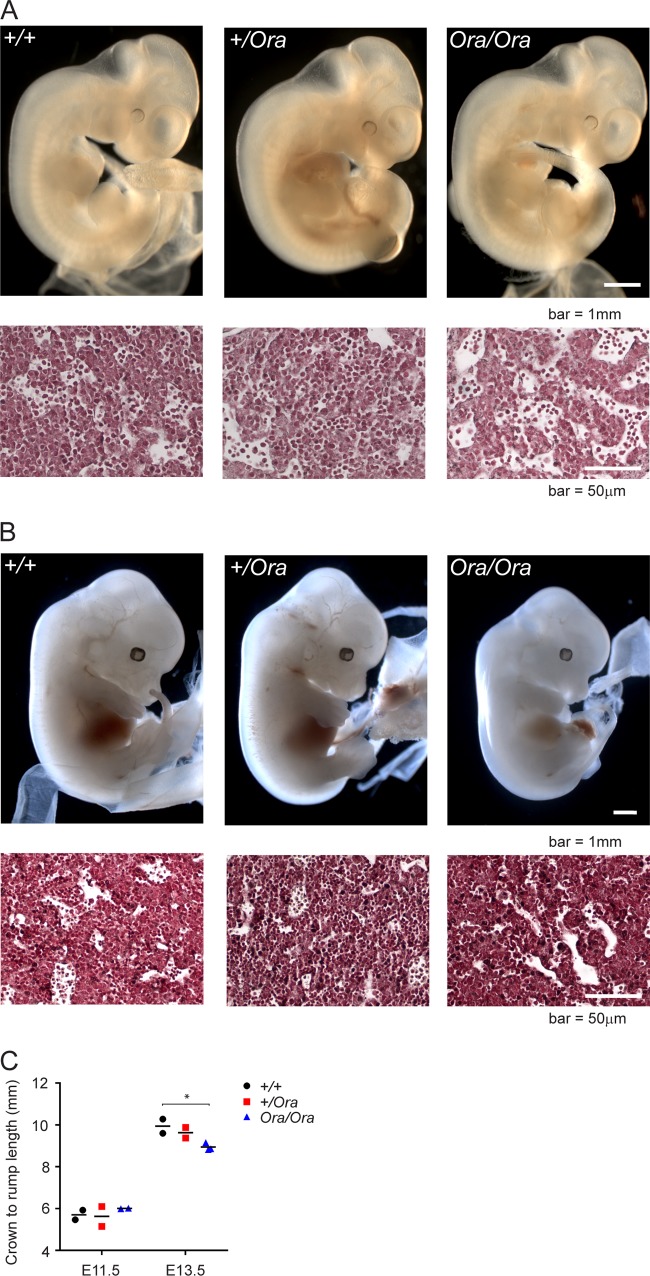
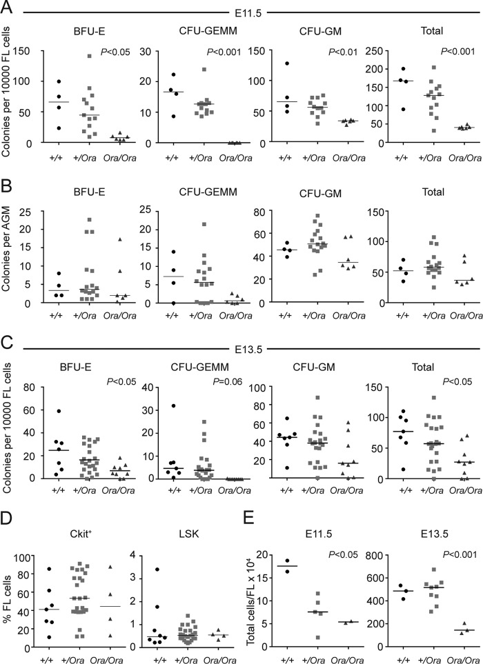
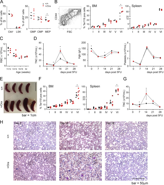
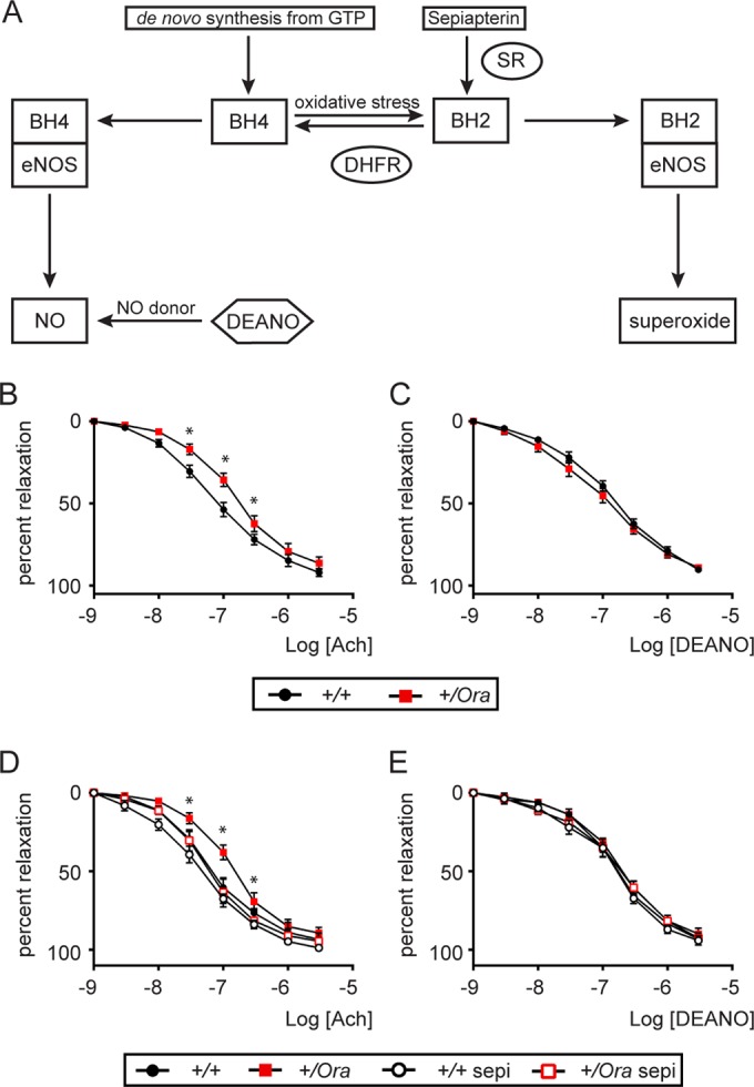
Dihydrofolate reductase (DHFR) is a critical enzyme in the folate metabolism pathway and also plays a role in regulating nitric oxide (NO) signaling in endothelial cells. Although both coding and noncoding mutations with phenotypic effects have been identified in the human DHFR gene, no mouse model is currently available to study the consequences of perturbing DHFR in vivo In order to identify genes involved in definitive hematopoiesis, we performed a forward genetic screen and produced a mouse line, here referred to as Orana, with a point mutation in the Dhfr locus leading to a Thr136Ala substitution in the DHFR protein. Homozygote Orana mice initiate definitive hematopoiesis, but expansion of progenitors in the fetal liver is compromised, and the animals die between embryonic day 13.5 (E13.5) and E14.5. Heterozygote Orana mice survive to adulthood but have tissue-specific alterations in folate abundance and distribution, perturbed stress erythropoiesis, and impaired endothelium-dependent relaxation of the aorta consistent with the role of DHFR in regulating NO signaling. Orana mice provide insight into the dual roles of DHFR and are a useful model for investigating the role of environmental and dietary factors in the context of vascular defects caused by altered NO signaling.


Figures

FIG 1
FIG 1
Orana mice have a point mutation in Dhfr, the gene encoding dihydrofolate reductase. (A) Schematic showing the ENU mutagenesis and screening strategy that led to the discovery of the Orana phenotype. The graph shows primitive (prim) RBCs and definitive (def) RBCs in blood from phenotypically normal (normal) and phenotypically mutant (mutant) E14.5 embryos. *, P < 0.001; t test. The bars in the graphs indicate the medians. (B) Sanger sequencing of +/+, +/Ora, and Ora/Ora mice showing a single A-G mutation at chromosome 13 base 92368224 (mm10). This mutation leads to an altered amino acid sequence of the DHFR protein (Thr136Ala). (C) Structure diagram (derived from Protein Data Bank [PDB] 4M6K [32]) showing human DHFR (gray) bound to folate (blue) and NADPH (not shown), indicating the position of Thr136 (red) in the folded protein. The dotted green lines indicate observed hydrogen bonds between Thr136, Glu30 (dark gray), water (green circle), and folate (30–32).
FIG 2
FIG 2
Mice harboring the Orana (Ora) allele have reduced levels of functional DHFR. (A) Relative mRNA expression in +/+, +/Ora, and Ora/Ora E13.5 fetal liver and in +/+ and +/Ora adult livers. (B) Protein expression in +/+, +/Ora, and Ora/Ora E13.5 fetal liver. The numbers indicate DHFR protein levels normalized to β-actin. (C) Relative DHFR protein expression in +/+ and +/Ora adult livers. (D) DHFR activity of equal amounts of protein from +/+ and +/Ora adult livers. (E) DHFR activity and Western blot of equal amounts of recombinant WT (DHFRWT) and Orana (DHFROra) DHFR protein expressed in E. coli. (F) Ectopic expression of WT and Orana DHFR in 293T cells before and after treatment with the proteasomal inhibitor BTZ. The bars in all the graphs indicate the medians.
FIG 3
FIG 3
Homozygous Orana embryos have defects in folate metabolism consistent with reduced DHFR activity. (A) Simplified flow chart of folate-mediated one-carbon metabolism illustrating the roles of different folate species. Dietary folic acid is reduced by the enzyme DHFR to DHF and THF. THF is converted to 5,10-methylene THF, an essential precursor for thymidylate synthesis, which is the rate-limiting step in DNA synthesis. 5,10-Methenyl THF and 5-formyl THF play important roles in purine synthesis. The recycling of 1 carbons back to THF occurs via 5-methyl THF, which is a critical folate species for methylation reactions, particularly DNA methylation. The consequences of a deficiency or imbalance of folate species are summarized. (B) Concentrations of folate species in +/+ (n = 3), +/Ora (n = 6), and Ora/Ora (n = 2) E13.5 whole embryos. P values are indicated (one-way analysis of variance [ANOVA]). (C) Overall abundance and distribution of folate species in E13.5 whole embryos. (D) DNA methylation levels in E13.5 whole embryos. (Left) Global. (Right) At individual CpGs adjacent to the Kcnq1ot1 promoter. The horizontal lines indicate mean methylation levels across all 10 CpGs for +/+ (black line; mean = 24.76%), +/Ora (red line; mean = 25.92%), and Ora/Ora (blue line; mean = 27.75%). The bars in all the plots indicate the medians.
FIG 4
FIG 4
Homozygous Orana FLs have defects in folate metabolism consistent with reduced DHFR activity, while adult heterozygotes have altered folate metabolism in a subset of tissues. (A) Concentrations of folate species in +/+ (n = 3), +/Ora (n = 6), and Ora/Ora (n = 2) E13.5 livers. The bars in the graphs indicate the medians. (B) Overall abundance and distribution of folate species in E13.5 livers. (C) Overall abundance and distribution of folate species in adult aorta, bone marrow, and liver, *, pregnant +/Ora mouse. (D) Global DNA methylation level in adult aorta, bone marrow, and liver. (E) Methylation of individual CpGs adjacent to the Kcnq1 promoter in adult aorta, bone marrow, and liver. The horizontal lines indicate mean methylation across all 10 CpGs for +/+ (black line; aorta mean = 27.43%, BM mean = 23.6%, and liver mean = 20.52%), +/Ora (red line; aorta mean = 26.11%, BM mean = 24.26%, and liver mean = 24.00%), and pregnant +/Ora (dashed red line; aorta mean = 24.97%, BM mean = 25.63%, and liver mean = 24.02%). The error bars indicate standard errors of the means (SEM).
FIG 5
FIG 5
Reduced DHFR function leads to altered embryonic morphology and reduced erythrocyte production in the fetal liver. (A) Whole mounts of +/+, +/Ora, and Ora/Ora E11.5 embryos and corresponding neutral-red-stained fetal liver sections. (B) Whole mounts of +/+, +/Ora, and Ora/Ora E13.5 embryos and corresponding neutral-red-stained fetal liver sections. (C) Crown-to-rump measurements of E11.5 and E13.5 embryos. *, P < 0.05 (t test). The bars in the graph indicate the medians.
FIG 6
FIG 6
Reduced DHFR function leads to defects in embryonic blood production. (A and B) Functional assays of blood progenitors in E11.5 FL (A) and AGM (B). P values are indicated (one-way ANOVA). (C) Functional assays of blood progenitors in E13.5 FLs. P values are indicated (one-way ANOVA). (D) Flow cytometry analysis of cKit+ and LSK cells in E13.5 FLs. (E) Total cells per FL at E11.5 and E13.5. P values are indicated (one-way ANOVA). The bars in the graphs indicate the medians.
FIG 7
FIG 7
Reduced DHFR function leads to alterations in the erythroid compartment and impaired response to 5FU-induced hematoablation. (A) Flow cytometry analysis of blood progenitors in adult bone marrow. *, P < 0.05 (t test). The bars in all the graphs indicate the medians; each data point indicates an individual mouse. (B) Representative flow cytometry plot indicating gates used to determine maturing erythroid populations in panels B and F. The graphs show relative abundances of Ter119+ erythroid populations in BM and spleens of +/+ and +/Ora adult mice. *, P < 0.05 (t test). (C) Peripheral blood RBC counts in young and older +/+ and +/Ora mice. (D) Plots showing total TNC, Hgb, and platelets in peripheral blood (PB) of 8- to 9-month-old +/+ and +/Ora mice following 5FU treatment. *, P < 0.05 (t test). (E) Spleens of 8- to 9-month-old +/+ and +/Ora mice 28 days after 5FU treatment. The asterisk indicates the spleen from mouse 503. (F) Relative abundances of Ter119+ erythroid populations in BM and spleens of 8- to 9-month-old +/+ and +/Ora adult mice 28 days after 5FU treatment. *, P < 0.05 (t test). The data points from the spleen of mouse 503 (red squares marked with ×) were excluded from statistical analysis. (G) Plots showing TNC of 2- to 3-month-old +/+ and +/Ora mice following 5FU treatment. (H) Wright-stained PB smears from 2- to 3-month-old +/+ and +/Ora mice following 5FU treatment. The arrows indicate abundant nucleated red cells in the peripheral blood. The error bars indicate SEM.
FIG 8
FIG 8
Reduced DHFR leads to impaired endothelium-dependent vasorelaxation in Orana mice, which can be restored by bolstering BH4 levels with sepiapterin. (A) Schematic representation of de novo BH4 synthesis and recycling pathways. BH4 is synthesized de novo from GTP via a series of reactions involving the rate-limiting enzyme GTPCH-1 and sepiapterin reductase (SR). BH4 is oxidized to BH2 in cells. DHFR can regenerate BH4 from BH2 via the recycling pathway. eNOS coupling is maintained when it is bound to BH4, producing NO, whereas BH2-bound eNOS leads to uncoupling and subsequent production of superoxide instead of NO. (B and C) Endothelium-dependent relaxation responses to Ach (B) and endothelium-independent relaxation responses to DEANO (C) of +/+ or +/Ora mice. The data are means ± SEM (n = 5). *, significant (P < 0.05) difference between +/+ and +/Ora aortic rings (unpaired t test). (D and E) Endothelium-dependent vasorelaxation responses to Ach (D) and endothelium-independent relaxation responses to DEANO (E) of +/+ or +/Ora mice in the absence or presence of sepiapterin (Sepi) (100 μmol/liter; 1 h preincubation). The data are means ± SEM (n = 3). *, significant (P < 0.05) difference between +/Ora and +/Ora Sepi aortic rings (unpaired t test). The relaxation responses to Ach or DEANO were recorded after vessel contraction with phenylephrine (300 nmol/liter).

Similar articles

Cited by

References

    1. Liu JJ, Ward RL. 2010. Folate and one-carbon metabolism and its impact on aberrant DNA methylation in cancer. Adv Genet 71:79–121. doi:10.1016/B978-0-12-380864-6.00004-3. - DOI - PubMed
    1. Thony B, Auerbach G, Blau N. 2000. Tetrahydrobiopterin biosynthesis, regeneration and functions. Biochem J 347:1–16. doi:10.1042/0264-6021:3470001. - DOI - PMC - PubMed
    1. Werner-Felmayer G, Golderer G, Werner ER. 2002. Tetrahydrobiopterin biosynthesis, utilization and pharmacological effects. Curr Drug Metab 3:159–173. doi:10.2174/1389200024605073. - DOI - PubMed
    1. Chalupsky K, Cai H. 2005. Endothelial dihydrofolate reductase: critical for nitric oxide bioavailability and role in angiotensin II uncoupling of endothelial nitric oxide synthase. Proc Natl Acad Sci U S A 102:9056–9061. doi:10.1073/pnas.0409594102. - DOI - PMC - PubMed
    1. Vasquez-Vivar J, Kalyanaraman B, Martasek P, Hogg N, Masters BS, Karoui H, Tordo P, Pritchard KA Jr. 1998. Superoxide generation by endothelial nitric oxide synthase: the influence of cofactors. Proc Natl Acad Sci U S A 95:9220–9225. doi:10.1073/pnas.95.16.9220. - DOI - PMC - PubMed

Publication types

MeSH terms

LinkOut - more resources